


Actin and Tubulin, as the major cytoskeleton structures, are crucial components of a plethora of processes in cell biology. Both are very much involved in stabilizing the cell shape, and especially in cell movements (e.g. cell migration) and intracellular movements and transport mechanisms.
Thus visualizing Actin and Tubulin in fixed or living cells and detecting them in biological samples (e.g. in Western Blots) belongs to basic experimental set-ups in Cell Biology.
A broad range of tools is available for all kind of experimental levels, in this post we’ll take a look at some of these you can use from Western Blot to Live Cell Imaging.
Detection of Actin and Tubulin with Antibodies

Antibodies can be used in diverse methods, such as Western Blots, ELISA, and Immunocytochemistry. Good results in different methods can be only obtained with a few antibodies.
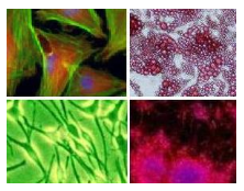
Anti pan Actin has been thoroughly tested in these application and brought brilliant results (See Fig 1 and 2).


In addition, here you can browse a selection of other anti Actin antibodies .
Anti-alpha/beta tubulin has been positively tested in the same applications and can additionally be used for Immunoprecipitation experiments. Results are presented in Fig. 3 and Fig. 4.
Here’s a complete overview of a range of anti Tubulin antibodies.
Actin staining of fixed cells

In fixed cells Actin can be stained with antibodies (see above) but often fluorescent phalloidins are used. Phalloidin belongs to the group of phallotoxins produced by the mushroom Amanita phalloides (death cap mushroom). The natural toxicity of Phalloidin is due to its stabilizing effect on F actin in cells. Based on its affinity for F actin and coupled to a fluorescent dye, it can be used to visualize F actin.
Cytoskeleton Inc. offers a set of phalloidin based stains (Acti-Stains) coupled to a number of different fluorophores compatible with popular filter sets such as FITC, TRITC and Cy5. The stains are exceptionally bright and stable and are indeed offered at very economical prices compared to other phalloidin based stains coupled to fluorophores of similar stability. Results of staining of Swiss 3T3 cells with ActiStain 488 are shown in Fig. 5.
Live Cell imaging of Actin and Tubulin

Finally, I would like to introduce brand new tools to stain Actin and Tubulin in living cells without the need to transfect them with vectors carrying the information for fluorescently labeled Actin/Tubulin or binding proteins. Spirochrome recently released unique fluorescent stains for monitoring both major components of thecytoskeleton easily in living cells. These fluorescent stains (SiR-Actin and SiR-Tubulin) are cell permeable compounds which stain F-actin (and microtubules, respectively; see Fig. 6 and Fig.7).

To learn more about these exciting stains, take a look at my recent post 2 new Actin and Tubulin live-cell imaging stains – without transfection!
Interested in staining Actin and/or Tubulin? Get in touch by leaving your questions or comments below!


3 responses
Hello and thank you all for this resource.
I have routinely used fluorescently conjugated phalloidin to stain actin cytoskeletal elements for years, in fixed, permeabilized cells.
Are there similar probes to be used as non-antibody stains of tubulin cytoskeletal elements in fixed, permeabilized cells?
Thank you very much.
Dear All,
Thank you for your question on our blog.
Our SIR tubulin probes are meant for live cell imaging.
I see you are based in the US. You may wish to discuss this futher with our partner Cytoskeleton Inc., based in the US.
They are actin and tubulin specialists.
Feel free to get in touch with me if you need guidance for this.
Here are my contact details: isabelle.nobiron@tebu-bio.com
Kind regards,
Isabelle
Did you ever find out? Please share if you have, thanks!!