Intracellular Flow Cytometry enables the identification and analysis of signaling and functional markers within cells from samples containing heterogeneous cell populations. Many cell types can be thus identified through their intracellular Flow Cytometry patterns and key intracellular signaling pathways and their respective post-translational modifications can be conveniently analysed. Behind these unique performances, Intracellular Flow Cytometry require anyhow optimized immunoreagents for cell permeabilization, intracellular staining and fluorescent detection.
4-stage Intracellular Staining for Flow Cytometry (source: Rockland Inc.)
Rockland immunochemicals, a US-based biotechnology company manufacturing specialized antibodies and assays, have recently described the basics of Intracellular Flow Cytometry in 4 steps:
- Cells are treated with drug or stimulant
- Cells are permeabilized and fixed
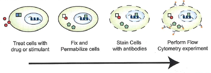
- Cells are stained with a fluorochrome conjuguated antibody
- Cells are ready for analysis of intracellular targets by flow cytometry
Fixation & Permeabilization for intracellular staining
Fixation and permeabilization of the cell membrane is a key step there. Once made, primary antibodies and fluorescent-conjugates can enter the cell and interact with their specific target antigen
The balance between intracellular target integrity and cell permeability might be optimal. According to Dr Camillo Moncada, Senior scientist & Director of QC at Rockland Immunochemicals, optimization of cell fixation and permeabilization is a critical parameter that needs to be empirically determined. For this, Camillo recommends to always use temporaneously freshly prepared solutions of high purity Paraformaldehyde for fixation and initially try mild detergents (i.e., Tween 20) for permeabilization.
If fixation and permeabilization steps require further optimisations, then gradual increasing formaldehyde concentrations (up to 4%) or ethanol or methanol and other detergents (including Triton X-100, or saponin without fixation or alcohol fixation) can be tested.
What about primary antibodies?
Primary antibodies are crucial too since they bring the specificity of the assay. It is strongly recommended to use primary antibodies compatible with flow cytometry and that have been initially validated to recognize post-translational modifications on key cell signaling pathway markers. A few examples:
- Anti -AKT1 PE: a mouse monoclonal antibody
- Clone 14E5.A2.B2.H9
- Applications: FCM, ELISA, , IHC, IF, WB
- Anti -AKT2 PE: a mouse monoclonal antibody
- Clone 16G11.E8 IgG2a kappa
- Applications: FCM, ELISA, , IHC, IF, WB
- Anti PPAR-ALPHA: a rabbit antibody
- Applications: FCM, ELISA, WB, IP
- Anti ATM Protein Kinase pS1981: a mouse monoclonal antibody
- Clone: 10H11.E12
- Applications: FCM, ELISA, Microscopy, WB
Interestingly, one of the most popular antibodies for intracellular detection by Flow Cytometry is related to the Green Fluorescent protein:
- Anti-GFP: a mouse monoclonal antibody
- Applications: FCM, ICC, IHC, ELISA, WB, IP
What about you, which are your favorite reagents and antibodies for Flow Cytometry of intracellular targets?