
Recently I reported about methods to measure:
- Collagen – Sircol for soluble and insoluble collagen assays
- Glycosaminoglycans – How to measure Glycosaminoglycans and Proteoglycans?
Today, I invite you to take a closer look at another component of the extacellular matrix (ECM) – Elastin.

Elastin is a highly elastic protein in connective tissue and enables tissues in the body to resume their shape after contracting or stretching. Elastin helps skin to return to its original position when it is pinched or poked. During aging, elastin appearance decreases. The ELN gene encodes a protein which is rich in hydrophobic amino acids like glycine and proline, which form mobile hydrophobic regions bounded by crosslinks between lysine residues. A number of transcript variants encoding different isoforms are know for this gene. Together with the elastic microfibril (consisting of proteins such as microfibrillar-associated glycoproteins, fibrillin, fibullin, and the elastin receptor) elastin forms so called elastic fibers in the ECM of connective tissues.
Elastin and Diseases
Deletions and mutations in the ELN gene are associated with Supravalvular aortic stenosis (SVAS) and the autosomal dominant cutis laxa (or Chalazoderma or Dermatochalasia). Further elastin-related defects include Marfan syndrome, emphysema, atherosclerosis, Buschke-Ollendorff syndrome, Menkes syndrome, pseudoxanthoma elasticum, and Williams syndrome.
Detection of Elastin
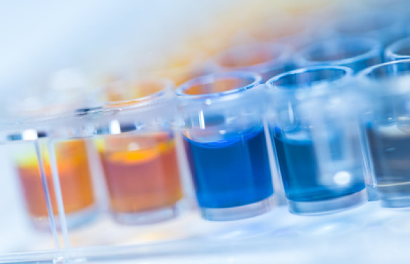
Often elastin is detected with either ELISA or immunohistochemistry based methods. Our partner Biocolor developed a quantitative dye-binding method for the analysis of elastins released into tissue culture medium and extracted from biological materials (see the Fastin Flowchart in Fig.1)

Which elastin forms can be measured by the Fastin Assay?
- soluble tropoelastins
- athyrogenic elastins
- insoluble elastins (following solubilization to elastin polypeptides [α-elastin, κ-elastin])
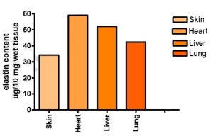
Fig. 2: Extraction of elastin from mouse tissue by hot oxalic acid digestion. The extracts shown were pooled and expressed as μg elastin per 10 mg wet tissue.


