

If the answer is yes, I am sure you’ll be interested to learn more about WST-8 and our Cell Counting Kit-8 (CCK-8). If the answer is no, and you’ve already switched to WST-8, just have a look at our price list… you might be surprised!
 CCK-8 allows sensitive colorimetric assays for the determination of cell viability in cell proliferation and cytotoxicity assays. Dojindo’s highly water-soluble tetrazolium salt, WST-8, is reduced by dehydrogenase activities in cells to give a yellow-color formazan dye, which is soluble in the tissue culture media. The amount of the formazan dye, generated by the activities of dehydrogenases in cells, is directly proportional to the number of living cells. The detection sensitivity of CCK-8 is higher than the other tetrazolium salts such as MTT, XTT, MTS or WST-1.
The 4 main benefits (to keep it short!)
- No toxicity to cells
- More sensitive than MTT, MTS, or WST-1
- 3 simple steps (no thawing necessary): Add – Incubate – Measure
- More stable than MTT, MTS or WST-1
Just keep on reading for more details about each of these benefits…
Cell Counting in 3 steps

The handling time of CCK-8 is the shortest compared to its competitors. Only 15 minutes of handling time is needed for CCK-8, whereas longer handling time is required for both MTS and MTT assays.

Unlike MTT assays, since there is no need to lyse the cells, you don’t have to worry about the data variation.
Stability
CCK-8 is a ready-to-use solution. It is also stable at 4ºC for 1 year. Your assay can be done anytime without thaw and freeze.

Sensitivity
CCK-8 is the highest sensitive dye for cell based assays.

Cytotoxicity
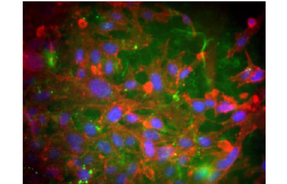
Only in CCK-8 is continuous culture possible without killing cells. Below are illustrations observing Cytotoxicity after incubation with each reagent. HeLa cells were incubated with WST-8 (CCK-8 ), WST-1, or MTS and observed at 24 hours after addition of these reagents.

Conclusion…
Well, since you’ve read this post right through to the end, I’m pleased to share this short cartoon with you!


6 Responses
Dear Sr/Srs.
We usually do cytotoxicity assays in the lab with XTT. It would be possible to get a free sample to prove in our lab the CCK-8?
Dear Aintzane,
I am pleased to hear that you are considering CCK-8 as an attractive alternative for your XTT cytotoxic assays. I am contacting you directly to define the modalities for the validation kit.
Looking forward hearing from you.
Regards,
Philippe
Dear Aintzane,
I’ll be glad if you can share your results briefly regarding WST-8 vs. XTT.
We use XTT in our lab, too. If you have better results with WST-8, I’ll think about buying WST-8.
Thanks in advance. Best regards,
Ezgi
Hello, I would like to know if you will answer this question, what is the method of CCK-8 to enter the cell?
Dear Jessie,
I would like to thank you for your interest in our post and products.
To answer your question regarding the CCK-8 product, one of the particularity of this new viability test is that the compound WST-8 is not entering into the cell (you can re use your cells after the viability analysis). The WST-8 compound stay outside the cell and it’s a ion acceptor who enter into the cell, and will stand out if the cell is living to reduce the WST-8 and produce orange colour. The intensity of the colour is directly proportional to the number of living cells.
You can find more information on the CCK-8 features in this link :http://5.196.212.231/2016/05/20/still-using-mtt-or-swt-1-for-cell-counting/
To allow you to try this powerful new viability assay, we can provide you a free sample to try. DOn’t hesitate to contact me directly (frederic.samazan@tebu-bio.com) if your are interested by this free testing sample.
Best regards,
Frédéric
Hello.
I would like to know if you have tested how is the background at low cell densities in this Kit and/or if you have tested its lower background at low cell densities compared to the MTS assay.
Thank you very much