

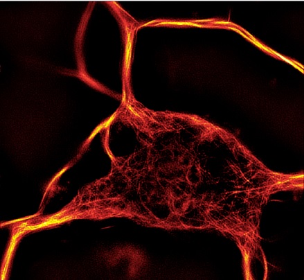
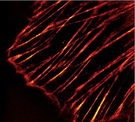
Cytoskeletal live cell imaging is extremely powerful when investigating cellular processes such as cytokinesis, motility and organelle transport and organization. The current experimental procedures remain nevertheless cumbersome and long. This post demonstrates how cell permeable, transfection free, Tubulin and Actin red fluorescent dyes help Cell biologists in analysing cytoskeleton dynamics in living cells.

Up to now the options for live cell staining of Actin and Tubulin molecules were limited. Phalloidin derivatives which selectively bind to F-actin are not cell permeable. They can thus only be used in fixed and permeabilized cells. Paclitaxel (Taxol) derivatives binding to microtubules and other cytoskeletal modeling molecules do not show increased fluorescence upon target binding (meaning they are not fluorogenic), anyhow they have a strong impact on tubulin dynamics. The only methods for live cell imaging available so far have been systems based on expression vectors which have to be transfected to target cells. In the case of Actin, a binding protein is coupled to either GFP or RFP and thus is expressed in target cells which leads to a labeling of F-actin. Such an experimental approach can bring major technical hurdles such as transfection efficiency and duration.
The need for “easy-to-use” and non toxic stains, which can pass the cell membrane and directly bind and stain microtubules or F-actin, has been a demand from cell biologists for quite some years now. Up to now phalloidine based dyes were recommended to stain F-actin, but these dyes – although being perfect tools to stain F-actin in fixed cells – cannot be used for living cells as already been mentioned above.
Non-toxic Actin & Tubulin fluorescent stains for live-cell imaging
Cell biologists at tebu-bio have selected Spirochrome’s unique fluorescent stains for monitoring Actin and Tubulin easily in living cells. These fluorescent stains (SiR-Actin and SiR-Tubulin) are cell permeable compounds which stain microtubules and F-actin respectively in living cells.
They are compatible with Super-Resolution Microscopy like Stimulated Emission Depletion [STED] and Structured Illumination Microscopy [SIM] and render live cell cytoskeleton dynamics studies convenient:
- No transfection
- No washing steps
- No toxic effects
- Excellent brightness
- Far-red excitation & emission
- Deep tissue penetration and minimal background
- Multiple fluorescent stainings with other dyes possible
Want to know more about these new SiR actin and Tubulin labelling probes?
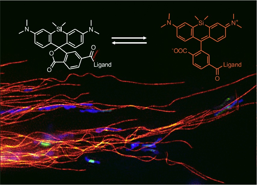
This proprietary bright and photostable silicon rhodamine-like (SiR) technology is compatible with most microscopes. It can be used with standard Cy5 settings. The combination of all these properties set SiR-based probes apart from other fluorescent probes. (1) The SiR dyes are coupled to binding components which specifically bind to F-actin (Jasplakinolide natural compound) and Microtubules (Docetaxel) in cells.
SiR derivatives exist in two states – the nonfluorescent spirolactone (OFF state) and a fluorescent zwitterion (ON state). Conjugated to Jasplakinolide or Docetaxel and with an optimized hydrophobic linker between the fluorophore and the targeting ligand, the final product SiR-Actin displays an increase in fluorescence of more than 100-fold upon binding to F-actin, while SiR-tubulin showed an increase of more than tenfold (2).
Sources:
- A near-infrared fluorophore for live-cell super-resolution microscopy of cellular proteins G. Lukinavičius et al., Nature Chemistry, 5, 132–139 (2013).
- Fluorogenic probes for live-cell imaging of the cytoskeleton, G. Lukinavičius et al., Nature Methods, 11, 731–733 (2014)
SiR-actin (Live Cell Fluorogenic F-actin Labelling Probe) and SiR-tubulin (Live Cell Fluorogenic Microtubule Labelling Probe) are available at 50 nmol separately. A complete Cytoskeleton kit with SiR-actin and SiR-tubulin (50 nmol each) is also available.
Leave your questions or comments here to learn more about these new cytoskeletal stains for fluorescent live cell imaging!

11 Responses
Hi,
I do actin/ß3-tubulin staining in growth cones of neurons and I am facing the problem that phalloidinOregongreen488 is immediately bleached out when exposing to STED laser (592 depletion). Also the signal intensity is not that great.
What are your experiences with SiR-Actin using STED.?
I would like to know it, because I have to use GA-fixation to preserve microtubule stability. As GA is not the best for acting due to the destruction of epitopes, I am wondering if I can use this SiR-actin and fix the cells afterwards with GA.
Thank you in advance.
Best regards
Dear Georg,
First of all, I would like to thank you for reading our blog and more specifically our article on the “2 new actin and tubulin live cell imaging dye”.
I’m pleased to confirm that the SiR-Actin is fully compatible with STED microscopy. You will find a series of publications displaying the use of SiR probes with STED here (http://5.196.212.231/2015/10/01/sir-dna_for_live_cell_imaging_of_dna_for_sted_and_sim/) or here (http://spirochrome.com/references/).
Regarding fixation, we’ve never had returns from glutaraldehyde fixed samples with SiR-probes. Nevertheless, it could advantageous to link the probe to actin and lead to a covalent linkage of the probe to F-actin (i.e. to fix with GA cells pre-stained with SiR-actin).
Unfortunately, we can’t make a stronger statement and guarantee the results as this has never been tested for the moment. Maybe if you are focusing only on targeting Actin, you can use PFA fixation instead as it mentioned in the FAQ of the SiR-Actin kit (http://spirochrome.com/product/sir-actin-50-nmol/).
I would like to mention too that SiR-tubulin and Sir-actin can be used in live cell imaging to reveal both networks.
I hope these information helps. Feel free to contact us for any further assistance.
Best regards,
Frédéric
I am interested in SiR-Actin and SiR-Tubulin. It appears that all successful cases or publications using SiR were on cell lines or cell culture. I would like to label actin in intact brain tissue, either acute brain slice preparations or in vivo. Do you know if anyone has tried this?
Dear Wan,
Thank you very much for your request and your interest for the SiR product.
As far as we know, the SiR-actin (or any other SiR probe) has not been used with in vitro brain tissue slices or in vivo. But there should be no problem that SiR-actin and SiR products will nicely work in brain slices.
Some exemple of SiR products use on tissue:
– SiR-DNA was used on fixed c.elegans.
– SiR-actin has also been used with snake skin samples.
– Sir probe used on mouse mammary gland sections.
Concerning the intact brain tissue the problem that may arise is the penetration of the probe deep into the tissue.
I will send you more information for the c-elegans used and the contact to SiR products US distributor by mail.
Best regards,
Fredéric
“– Sir probe used on mouse mammary gland sections.”
Do you have a link to the article?
Best regards
Olaf
Dear Olaf,
First of all I would like to thank you regarding your interest for our blog and also the products distributed by tebu-bio.
More specifically, I confirm you that SiR probes are working on mouse mammary gland sections. Unfoertunately this information is currently not published and the results came out through a technical discussion with our supplier.
Maybe we can discuss more in details about your experiments and find the best protocol?
Don’t hesitate to contact direclty by mail: frederic.samazan@tebu-bio.com
Best regards,
Frédéric
I am looking for a far-red tubulin staining for tubulin dynamics during live cell imaging that can be excited with a 640nm laser. Is your dye suitable? Thanks a lot.
Dear Yvonne,
The 640 nm laser line is just perfect. Interestingly, even a 633 nm laser line works also.
Regards,
Philippe
Hi,
I would like to use SiR-Actin and SiR-Tubulin in 3D tissue constructs with fibroblasts and do the live cell imaging. It doesn’t look like anyone tried it in a 3D tissue environment, just on plated cells. Do you know if anyone has tried this?
Regards,
Dela
Dear Dr Shakiba,
FIrst of all I would like to thank you regarding your interest for our posts and products.
Regarding your specific request I confirm you that customer already used the SiR- Actin on Human Colon Organoids for live cell staining.
Please don’t hesitate to contact me directly to discuss more in details about this protocol for the use of SiR probes on 3D culture.
Best regards,
Frédéric
frederic.samazan@tebu-bio.com
+33 1 30 46 39 86
Hi everybody,
I would like to use the Actin and Tubulin live cell imaging staining on T blasts using the spinning disk at 63X. The idea is to label those two structures and make cells migrate through channels. Would like to see labeled structures in live cells. Would it be possible ?