According to the WHO (World Health Organization), early diagnosis of diseases is one of the key factors in the world to save lives and reduce the cost of treatment. Faced with these recommendations, in vitro diagnostic tests can prevent, detect, diagnose, evolve, monitor, and manage a pathology, but also select and follow a therapy. Current in vitro tests are designed biological products coupled with fluorescent molecules to detect biomarkers, and thus diseases. These dyes emit a signal that can be observed and quantified and represent one of the central elements to diagnose pathology.
Nowadays, the detection of biomarkers is addressed by several products developed in the 2000s whose performance is moderate. Fortunately, a lot of research and many technologies are appearing to push the limits of the detection of biomarkers, such as lanthanide nanoparticles technology.
What is unique about lanthanide nanoparticles?
Lanthanides are chemical elements having the atomic number Z from 57 to 71. They are considered “rare earth” and their general electronic configuration is [Xe] 4fn5d16s2 where n is included between 0 to 14 and represents the number of electrons and [Xe] corresponds to the electronic configuration of the noble gas xenon.
Each lanthanide elements have different optical and magnetic properties and are mainly used as catalysts, magnets, optical lasers, or fibers but their specific properties have also attracted life science and medical areas, especially for the detection of biomarkers. With a large energy gap between the lowest excited state and the highest ground level, each lanthanide has a specific spectroscopic signature. The spectral range of lanthanides is large and they emit a strong luminescence in the visible region (Sm3+, Eu3+, Tb3+, Dy3+) or in the near-infrared region (Nd3+, Er3+, Ho3+, Tm3+, Yb3+).

Figure 1: Spectral range of lanthanides.
On the other hand, the power of nanoparticles resides in their nanometric structure. Nanoparticles are ultra-small particles, between 1 to 100 nm, and are used in many sectors such as healthcare, environment preservation, or the cosmetic industry. Nanoparticles present many advantages like small size, large surface area for functionalization, and good stability.
The combination of these two elements, nanoparticles, and lanthanides, lead to exceptional performance for the detection of biomarkers.

Figure 2: Bright-Dtech™ advantages.
How can they upgrade the detection of biomarkers?
Thanks to their properties, lanthanide nanoparticles represent a real revolution in the detection of biomarkers, allowing more precise detection and quantification thanks to their high sensitivity and specificity.

Figure 3: Revolution in the detection of biomarkers.
Indeed, these lanthanide nanoparticles have a long emission lifetime that can reach some milliseconds for Terbium and Europium, high brightness, high photostability and remove background noise for various applications in a better working time.

Figure 4: Bright-Dtech™ technology.
As current fluorescent markers, they can be used in several applications such as:
- TR-FRET
- TR-FLISA
- Lateral Flow (Rapid test)
- Multiplexing
- Western Blot
- In vitro applications
- Immunofluorescent for microscopic analysis
- Quantitative PCR

Figure 5: How it works?
The use of lanthanide nanoparticles in immunoassays represents a revolutionary step forward in the early detection and diagnosis of diseases such as cancer. Their exceptional sensitivity, specificity, and multiplexing capabilities have the potential to transform diagnostics, leading to earlier interventions and improved patient outcomes.
Interested in the use of lanthanide nanoparticles for your assays?
Find out more about lanthanide nanoparticles for your immunoassays and get access to our partner POLY-DTECH’s innovative technology to push forward your research.
Learn more
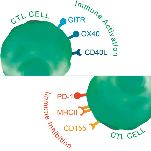
Your complete tool box to facilitate your Immunotherapy research
References
- Bünzli JC, Piguet C. Taking advantage of luminescent lanthanide ions. Chem Soc Rev. 2005;34(12):1048-1077. doi:10.1039/b406082m
- Gálico DA, Santos Calado CM, Murugesu M. Lanthanide molecular cluster-aggregates as the next generation of optical materials. Chem Sci. 2023;14(22):5827-5841. Published 2023 May 10. doi:10.1039/d3sc01088k
- Cheignon C, Kassir AA, Soro LK, Charbonnière LJ. Dye-sensitized lanthanide containing nanoparticles for luminescence based applications. Nanoscale. 2022;14(38):13915-13949. Published 2022 Oct 6. doi:10.1039/d1nr06464a
- Goetz J, Nonat A, Diallo A, et al. Ultrabright Lanthanide Nanoparticles. Chempluschem. 2016;81(6):497. doi:10.1002/cplu.201600117
- Charpentier C, Cifliku V, Goetz J, et al. Ultrabright Terbium Nanoparticles for FRET Biosensing and in Situ Imaging of Epidermal Growth Factor Receptors*. Chemistry. 2020;26(64):14602-14611. doi:10.1002/chem.202002007
Acknowledgment
Special thanks to Ms. Justine Renaud, intern at POLY-DTECH, for writing this article.