Stimulated emission depletion (STED) microscopy is an accurate process allowing super resolutive cell imaging. STED microscopy overcomes the limits of standard confocal laser scanning microscopes. In 2014, Stefan W. Hell was awarded the Nobel Prize in Chemistry, along with Eric Betzig and William Moerner (USA) for the development of this breakthrough technique. STED offers exceptional experimental conditions for visualization of cellular events at unprecedented levels. These levels can only be reached with high quality reagents. Here, let’s review some top quality research antibodies and fluorochromes validated for STED applications, and browse a gallery with outstanding cell images.
High quality research antibodies, together with the use of the next generation fluorochrome conjugates, offer superior absorption and thus, high fluorescence quantum yield and superior brightness & contrast. This post displays images obtained in collaboration with Rockland Immunochemicals and Leica Microsystems. The images presented here were obtained with the following experimental conditions:
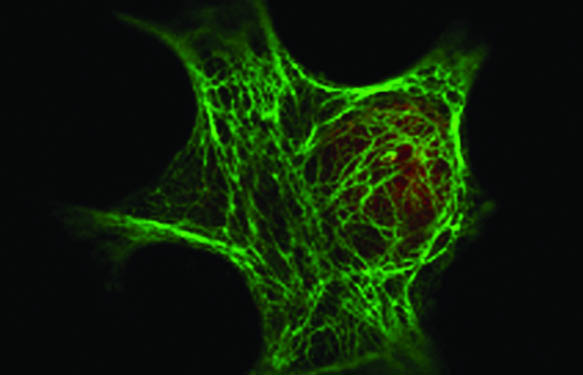
Epidermoid carcinoma cell line A431 stained for HDAC1 (Anti-HDAC1, 039600-401-879 + anti-Rabbit IgG ATTO 550, 039611-154-122) (red) and Keratin (Anti-Keratin, 039200-301-390 + anti-Mouse IgG DyLight 488, 039610-141-002)(green).
HDAC1 localizes at the nucleolar periphery.
The cell was imaged using a TCS SP8 STED 3X microscope (Leica Microsystems, Inc.) using a white light laser tuned to 470nm to excite the DyLight 488 fluorescence and 550nm to excite the ATTO 550.
For STED images, DyLight 488 was used with the 592nm depletion laser set to 95% power with a time gating range of 1.5-6.0 ns, and ATTO 550 was used with the 660nm depletion laser set to 100% power with a time gating range of 1.5-6.0 ns.
Galery of images obtained with the STED microscopy (source: Rockland Immunochemicals – Leica Micosystem)


Looking for research reagents compatible STED Microscopy?
High resolution can only be reached with robust and validated research reagents and protocols. This level of expertise is available in the States at Rockland Immunochemicals and in Europe through tebu-bio. If you’d like to know more about STED microscopy-compatible antibodies and Immuno-assays, leave a message or comment in the form below!