In the field of neurosciences, increasing research is being undertaken on neurodegenerative disorders like Parkinson’s and Alzheimer’s diseases, as well as on nerve injury and regeneration. One of the challenges researchers face is finding the appropriate cellular neuromodels, as close as possible to the in vivo environment. Primary cells are one of the answers, increasingly used in co-culture systems and 3D models. However, cells from the nervous system are not readily accessible, nor are they easy to culture.
Cell Applications Inc. provide solutions to face these challenges, ranging from highly pure, low passage ready-to-use primary cells, to stem cells and induced pluripotent stem cells (iPSC) derived from a variety of clinically relevant species. Let’s take a look at a selection of their neuron, microglia and astrocyte models in relation to their potential applications.
Human models
In an area where primary cell sourcing and culture is a challenge, Stem cells and iPSC appear as extremely interesting alternatives.
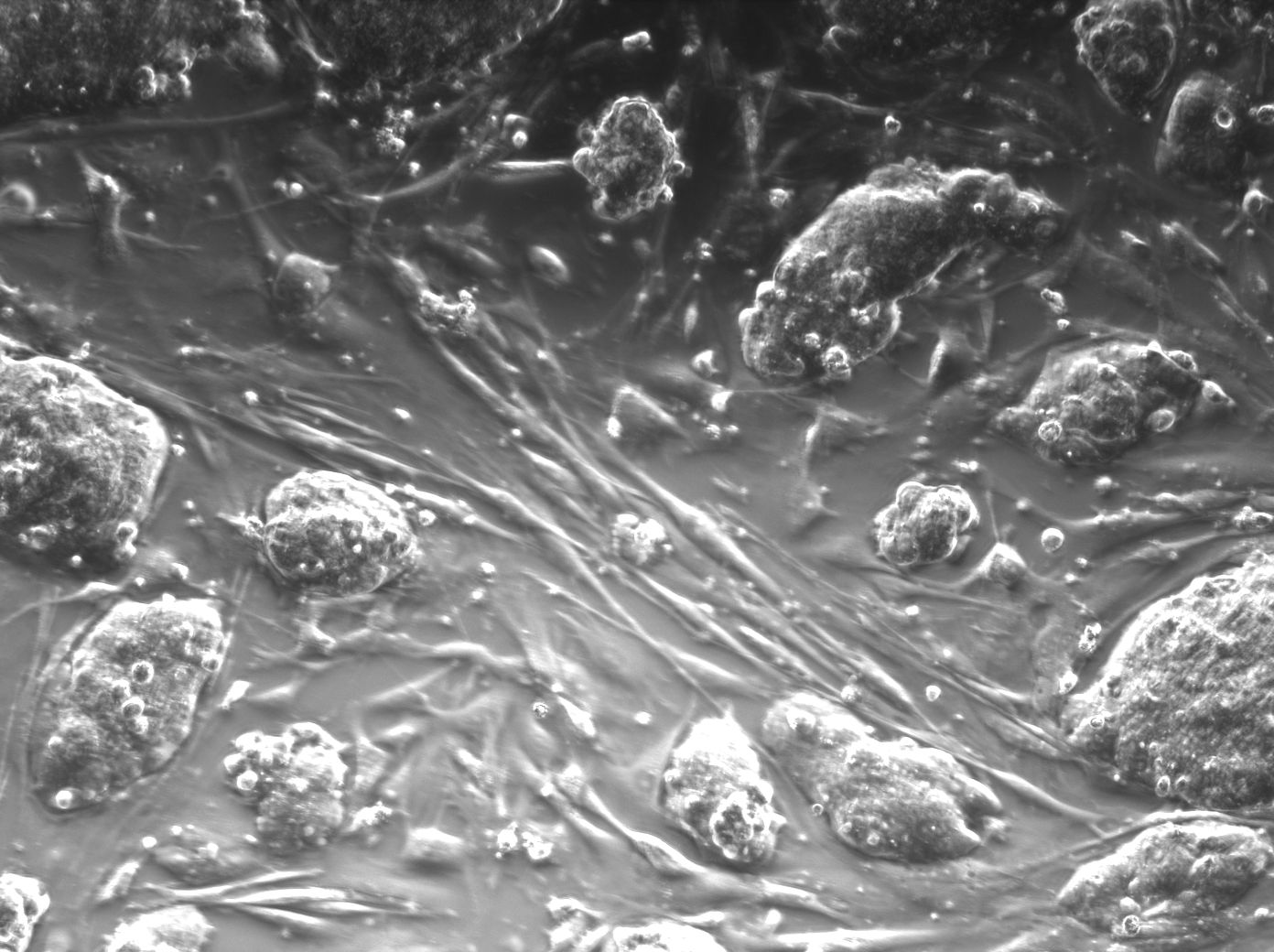
Human Neural Stem Cells (HNSC) are self-renewing, generated throughout an adult’s life via neurogenesis. These multipotent adult stem cells generate the main phenotype of the nervous system, differentiating into neurons, astrocytes, and oligodendrocytes. HNSC play important roles for development, learning and hippocampal plasticity. They are also used to study age-related declines in proliferation, as well as neurological diseases like stroke, multiple sclerosis, and Parkinson’s disease. The cells respond to injury, and can be differentiated to replace lost or injured neurons. They migrate in a directed fashion to brain tumours and help replace dying neurons in injured adult brain tissue.

Cell Applications HNSC are primary cells derived from the cortex region of human brain (single donor). They are cryopreserved at first passage and stain positive for β-tubulin III, GFAP and oligodendrocyte marker O4 when cultured in Human Neural Differentiation Medium for 10 days. Each lot is tested for its ability to form neurospheres in Human Neural Stem Cell Growth Medium.
 Human iPSC-Derived Neural Stem Cells (i-HNSC) are a homogeneous, self-renewing and multipotent population derived from Human Induced Pluripotent Stem Cells (HiPSC). Under controlled conditions, i-HNSC possess the ability to consistently generate high yields of functional neural cells like neurons and glia. i-HNSC express typical markers of neural stem and progenitor cells such as Nestin and Sox1 with a purity greater than 90%. They are completely validated for viability, differentiation potential, karyotype, morphology, passage number, and sterility. These i-HNSC provide researchers a unique model to study neural development, neurotoxicity, differentiation, electrophysiology, and disease modeling. i-HNSC or i-HNSC-derived neurons are useful for drug screening and research for cell-based therapy applications.
Neuronal Multi-electrode arrays (MEA) measure real-time network activity of cultured human neurons. The MEA measurements video below illustrates in i-HNSC-derived neurons the presence of a mature, synchronized neuronal network with optimal electrophysiological activity, which can be modulated by neurotransmitters or small compounds.
Human astrocytes (HA) are derived from human cerebral cortex. Astrocytes are the most abundant cells in the central nervous system where they perform many functions, such as providing mechanical support and nutrients to neurons and removal of wastes from neurons; providing signaling to endothelial cells; regulating neurogenesis and controlling synaptic function. As the recognition of the importance of astrocytes in nervous system is increasing, HA serve as a useful in vitro model for exploring the diversity of astrocytes functions, especially in glioblastoma research.

Cell Applications astrocytes are derived from normal healthy human brain; they are able to attached, spread and proliferate in growth medium. Provided at passage 3, a minimum of 10 doublings is guaranteed.
Rat models
 Rat Cortical and Hippocampal Neurons facilitate discovery for memory and age-related CNS disorders like Alzheimer’s disease, respectively. Rat Dorsal Root Ganglion Neurons help examine nerve injury and regeneration, while those working on coordination and equilibrium select Rat Hindbrain Neurons. Similarly, Rat Midbrain Neurons play roles in muscle control, Parkinson’s and ALS. Progress continues in spinal cord disorders like Friedreich’s ataxia, suitable for study in Rat Spinal Cord Neurons, while Rat Striatal Neurons provide insights into Parkinson’s.
 Microglia, originated from the hematopoietic stem cells in bone marrow, are the resident macrophages in the central nervous system (CNS) which is separated from the rest of the body due to the presence of blood-brain barrier. Microglia actively survey the surrounding area and respond by scavenging damaged neural cells, plaques, and infectious agents. Due to their functions in immune response and maintaining homeostasis in the CNS, microglia have been implicated in neurodevelopment, CNS plasticity and repair, neuroinflammation, aging and neurodegeneration, neuropathic pain, and infections.
Cell Applications rat microglia (RMcg) are derived from the brain of normal postnatal day 1 rats by standardized methods. When revived and plated and cultured under recommended conditions, RMcg form adherent culture.
A summary of the full rat neuro-models is illustrated below:

Rat Cortical Neurons, Rat Hippocampal Neurons, Rat Dorsal Root Ganglion Neurons, Rat Striatal Neurons, Rat Spinal Cord Neurons, Rat Astrocytes

Rat Midbrain Neurons, Rat Hindbrain Neurons, Rat Schwann Cells, Rat Perineurial Fibroblasts, Rat Microglial Cells, Rat Brain Microvascular Endothelial Cells
This is only a selection of the neuromodels available. Contact me if you can’t find the one you are looking for and I’ll be happy to help, also if you’re looking for live cell imaging tools or neuro cell lines.