Correlation of findings in animal models (e.g. mouse) and its validation in human samples lies at the basis of Translational medicine. Nowadays, hypothesis established in mouse models must, at some stage, be validated in a cohort of patients.

A paper by Ichii, S. et al. is a good example of this. They studied the role of miRNAs in the pathogenesis of autoimmune glomerulonephritis.
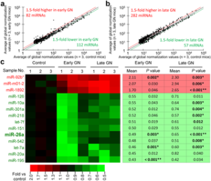
In mouse models (B6.MRLc1), after performing a miRNA profiling using Toray’s 3D Technology, they found that miR-26a was the most abundantly expressed microRNA in the glomerulus of normal C57BL/6 and that its glomerular expression in B6.MRLc1 was significantly lower than that in C57BL/6.
In mouse kidneys, podocytes mainly expressed miR-26a, and glomerular miR-26a expression in B6.MRLc1 mice correlated negatively with the urinary albumin levels and podocyte-specific gene expression. Puromycin-induced injury of immortalized mouse podocytes decreased miR-26a expression, perturbed the actin cytoskeleton, and increased the release of exosomes containing miR-26a.
They studied then patients with lupus nephritis and IgA nephropathy, and found that glomerular miR-26a levels were significantly lower than those of healthy controls. In B6.MRLc1 and patients with lupus nephritis, miR-26a levels in urinary exosomes were significantly higher compared with those for the respective healthy control.
These data indicate that miR-26a regulates podocyte differentiation and cytoskeletal integrity, and its altered levels in glomerulus and urine may serve as a marker of injured podocytes in autoimmune glomerulonephritis.
Want to translate your results in animal models to human samples? Leave your comment!


