As scientists better understand the benefits of growing cells in three dimensions (3D) and routinely adopt 3D culture techniques, methods for visualising cells must also be adapted and optimised.
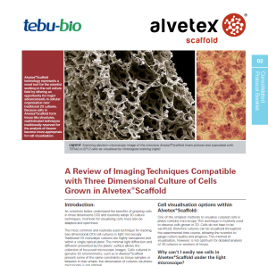
 The most common and routinely used technique for tracking two dimensional (2D) cell cultures is light microscopy. Traditional 2D monolayer cultures are highly transparent and within a single optical plane. The minimal light diffraction and diffusion presented by the plastic surface allows the collection of focussed microscopic images. Cells cultured in genuine 3D environments, such as in Alvetex®Scaffold, present some of the same constraints as tissue samples or biopsies, in that simple, live observation of cultures via phase microscopy is not optimal.
There are however, other techniques that can be implemented which will allow the user to monitor culture progress easily and effectively in 3D.Simple dyes can be used to identify culture confluence and viability. The variety of end-point visualisation techniques available to those culturing cells in 3D is extensive. Options include, but are not limited to, live cell imaging, fluorescent marker analysis, confocal analysis, histology using a range of cytological stains and electron microscopy. All of these techniques have been performed on cultures grown in Alvetex®Scaffold with excellent results.
To visualise cells cultured in Alvetex®Scaffold, a range of appropriate methods are available.
These are discussed in “A Review of Imaging Techniques Compatible with Three Dimensional Culture of Cells Grown in Alvetex®Scaffold”.
Request your free copy of this review here!
Related readings
- Formation of Mesenchymal Tissues in Alvetex® Scaffold
- Case study: 3D Cancer Cell Cytotoxicity in vitro Assessment
- A polystyrene scaffold for 3D endocervix model
- Alvetex on the International Space Station!