
Visualizing fixed cells and tissues only gives snap shots of cellular processes. Over the past years, more and more cellular parameters have become measurable in living cells. For this purpose, there is a growing number of specific stains capable of entering the cell without toxic effects (which could also negatively impact the parameters to be measured).

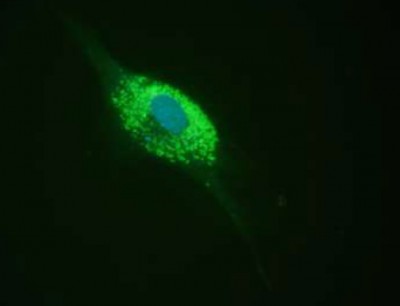
Organelles are specialized and well-defined parts of the cell which are usually separately enclosed within their own lipid bilayers. Very often organelles have to be stained to follow cellular processes and the effects of changing parameters on these processes. Thus there is a strong need for tools to stain and follow specific organelles in living cells.
An attractive offer of tools is available for this purpose – take a few moments to browse the respective selections related to the keywords below:

To get a complete overview of all available live cell imaging stains (actin, tubulin, diverse ions, intracellular pH, temperature etc.), you might be interested in consulting this interactive live cell imaging page.