Due to its involvement in many cellular processes such as normal ageing, tumor suppression, chronic diseases and many more, cellular senescence has gained increasing attention since its first description over 50 years ago. In addition, understanding of cellular senescence is invaluable in the development of therapeutic targets in the fight against age-related co-morbidities, cancer and chronic disease progression.
Over-expressed in senescent cells, Senescence-associated β-galactosidase (SA-β-gal), has been widely used as a marker of cellular senescence. In order to specifically detect this biomarker directly in living cells or tissues, let’s take a look at an easy and ready-to-use assay recently developed: the SPiDER-βGal- Cellular Senescence Detection kit.
SPiDER-βGal- Cellular Senescence Assay: principle
Based on the use of the new Dojindo Technology’s SPiDER-β Gal compound, this assay will detect SA-β-gal with high sensitivity and easily in only 30 minutes. Thanks to high cell-permeability and high retention inside cells, SPiDER-β Gal can specifically detect SA-β-gal in living cells by using a reagent, Bafilomycin A1, to inhibit endogeneous β-galactosidase activity.

In addition, SA-β-gal in fixed cells is also detectable by using McIlvaine buffer (pH 6.0). Since SPiDER-βGal emits strong and stable fluorescence after the reaction with SA-β-gal, it can be applied for fluorescent microscopy detection and also to quantitative analysis by flow cytometry.

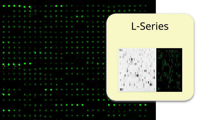
A. Passage 0, B. Passage 12
(green: SPiDER-βGal, blue: Hoechst 33342)
(Ex:500 ~ 540 nm)
(Em:530 ~ 570 nm)

positive WI-38 cell
Advantages against classical X-Gal detection
Although X-gal is a well-known reagent to detect SA-β-gal, it shows several disadvantages in comparison to the new SPiDER-βGal- Cellular Senescence Detection kit.
- Requirement of fixed cells due to the poor cell-permeability,
- Low quantitative capability because of the difficulty of the determination of visual difference between stained cells and not stained cells,

- Requirement of a long time of staining.

Whereas X-Gal method requires visual observation, the SPiDER-βGal method enables easier quantification of SA-β-Gal since it is applicable for Flow cytometry as well as Fluorescent microscopy.
Interested to use this new Cellular Senescence Assay ?
The SPiDER-βGal- Cellular Senescence Detection kit is available throughout Europe at tebu-bio’s web site.
You might also be interested in other Live Cell Imaging probes to detect senescence in High Content Screening assays, such as :
Contact us for more information via the form below, we’ll be pleased to advise!