Actin serves as one of the major cytoskeleton structures. It is a crucial component involved in a plethora of processes in cell biology: stabilizing the cell shape, cell movements (e.g. cell migration) and intracellular movements and transport mechanisms.
Actin is a 43 kDa protein that is very highly conserved between species. Actin has three main isotypes (α-actin, β-actin and γ-actin), which show >90% amino-acid (aa) homology between isotypes and >98% homology within members of a particular isotypic group.
A brief reminder: G-actin polymerizes to form F-actin

Globular-actin (G-actin) readily polymerizes under physiological conditions to form Filamentous-actin (F-actin) with the concomitant hydrolysis of ATP. F-actin is a double-helical filament (Fig. 1). Actin can polymerize from both ends in vitro. However, the rate of polymerization is not equal. This results in an intrinsic polarity in the actin filament. It has therefore become the convention to term the rapidly polymerizing end the plus-end or barbed-end (+) while the slow growing end is called the minus-end or pointed-end (-).
In the coming weeks I will give you an overview about methods in actin research with validated R&D products and kits (actin binding and actin binding proteins, actin polymerisation, and G-F actin ratio detection in cells).
Today I will focus on methods to visualize actin in fixed or living cells – which belong to basic experimental set-ups in Cell Biology.
Actin staining of fixed cells

Often fluorescent phalloidins are used to stain actin in fixed cells. Phalloidin belongs to the group of phallotoxins produced by the mushroom Amanita phalloides (death cap mushroom). The natural toxicity of Phalloidin is due to its stabilizing effect on F actin in cells. Based on its affinity for F-actin and coupled to a fluorescent dye, it can be used to visualize F-actin.
Cytoskeleton Inc. offers a set of phalloidin based stains (Acti-Stains) coupled to a number of different fluorophores compatible with popular filter sets such as FITC, TRITC and Cy5. The stains are exceptionally bright and stable and are indeed offered at very economical prices compared to other phalloidin based stains coupled to fluorophores of similar stability.
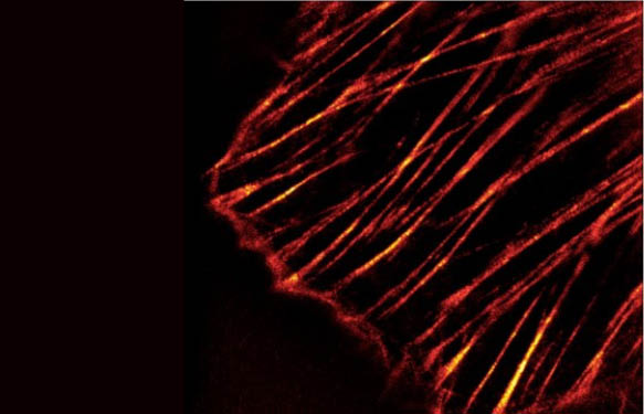
Results of staining of Swiss 3T3 cells with ActiStain 488 are shown in Fig. 2.
Live-cell imaging of Actin
Live-cell imaging of actin has been quite tricky as far as actin labelling is concerned – either cells had to be transfected with vectors carrying the genetic information for fluorescently tagged actin or actin binding proteins or, labelled actin had to be micro-injected to single cells.

Together with Spirochrome, tebu-bio launched in Europe the first tool to directly label actin in living cells with no need to transfect or micro-inject anything.
SiR-Actin is a cell permeable compound which stains F-actin in living cells. The stain is composed of a photostable silicon rhodamine-like (SiR) dye which can be used with standard Cy5 settings and a component (Jasplakinolide) which specifically binds to F-actin (Fig. 3). SiR-actin is compatible with Super-Resolution Microscopy like Stimulated Emission Depletion [STED] and Structured Illumination Microscopy [SIM].
f you would like to get an overview about the results SiR-actin users got so far, please have a look at my recent blog: User experience of SiR-Actin and SiR-Tubulin Live Cell Imaging.
Interested in our Phalloidin and/or SiR-based stains?
Leave your comment or request in the form below.