With their polycationic properties, polyamine organic compounds are involved in various biological functions (DNA-RNA interaction, gene expression…), and can also be considered as cancer marker.
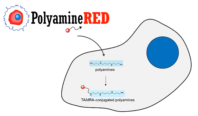
In this post, I’m pleased to introduce the first intra-cellular polyamine imaging reagent which works without any pre-treatment or cell lysis. This innovative reagent can be used with fixed cells, thus making it suitable for detection or semi-quantitative analysis of intra-cellular polyamines.
Background of Polyamines
Polyamines, such as putrescine, spermidine and spermine and its acetyl derivatives, are some of the essential classes of metabolites which have liner alkyl structure with two or more amines. As mentioned before, due to their polycationic properties, they show various biological functions. Polyamines also interact with negatively charged proteins and control their function. The major source of polyamines is amino acid ornithine. In the case of mammalians, ornithine is converted to putrescine by ornithine decarboxylase (ODC), followed by synthesizing spermindine and spermine. As ODC is highly expressed in cancer cells, polyamines are considered as cancer markers.
Several detection methods of polyamines have been developed so far, but most of them are low-throughput systems using HPLC with polyamine standard compounds. Therefore, biological functions of polyamines have been unclear. To answer this problem, the Polyamine RED (#FDV-0020) has been developped by Funakoshi.
Features of PolyamineRED (Cat. nr. FDV-0020)

PolyamineRED (cat. nr. FDV-0020) is the world’s first reagent for detecting intra-cellular polyamines without any pre-treatment and cell lysis.
This TAMRA (tetramethylrhodamine)-conjugated derivative of glycine propagyl ester specifically reacts with linear primary alkylamine and has cell-penetrating properties. It specifically reacts with polyamines inside the cells and labels polyamines with red fluorescent dye TAMRA (Fig. 1).
PolyamineRED : Specific features
- Specifically labels polyamines
- Can be detected by filter set for Rhodamine (Ex./Em. = 560 / 585 nm)
- Can be only used with fixed cells.
- No pre-treatment required
- Can do semi-quantification of total polyamines amount (*) by intra-cellular fluorescence intensity.
* Only total polyamines, not for each polyamine.
PolyamineRED : Chemical Information
- Molecular weight : 611 g/mol
- Solubility : Soluble in DMSO
- Fluorophore : TAMRA (red fluorescent dye)
Data obtained with PolyamineRED
Take a look below at some experimental data obtained with the PolyamineRED reagents.

In the figure 2, three cancer cell lines (MCF7, MDA-MB-231 and SK-BR-3) and two non-cancer cells (MCF10A and human lymphocyte) were treated with 30 μM of PolyamineRED for 10 min. After incubation, cells were washed three times by PBS, followed by DAPI staining and formalin fixation. Images were obtained at Ex/Em=560 nm/585 nm for TAMRA and at Ex/Em=358 nm/461 nm for DAPI. Finally TAMRA fluorescence was detected in cancer cell lines, showing a specific labeling of polyamine.

Figure 3, MDA-MB-231 cells were treated with 30 μMof PolyamineRED for 10 min. After incubation, cells were washed three times by PBS, followed by DAPI-staining and formalin fixation. Images were obtained at Ex/Em=560 nm/585 nm for TAMRA and at Ex/Em=358 nm/461 nm for DAPI. Major TAMRA signal was detected in nucleus. This indicates polyamines in MDA-MB-231 are mainly localized in nucleus.

Benzyloxycarbonyl glycine propagyl ester as a model molecule was selectively reacted with polyamines. Reactant of epinephrine, an example of monoamine, and lysine, an example of amino acid, were rarely detected. Reactivity for polyamines depends on the length of polyamine and double linkage products were observed from spermine (4 amino groups) and spermidine (3 amino groups) (Fig.4).
If you’re interested and want more information on this innovative reagent you can download the flyer, visit our web site or directly get in contact with your local tebu-bio office.

Interested in discovering other innovative tools for Live Cell Imaging?
Check out all our solutions on our interactive Live Cell Imaging Tools web page.