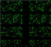
One of the questions we usually get in the Biomarkers team, once researchers or clinicians have done profiling arrays (e.g. for secretome and kinome), is how to interpret, biologically speaking, the obtained data.
It’s not the scope of this post to give a general overview of what has been published so far, but you can always have a look at publications using profiling arrays to see how other people have presented their results. The aim of this post is to give some general “rules” or ideas to help you analyse your data… even before you do your experiment.
Tip #1 – identity of samples
A good profiling experiment (as any experiment) starts with the right choice of the samples to be analysed. You may decide to use individual samples, or to pool them. This depends on your project and what you intend to obtain.
As a rule of thumb, in our team, we normally advise that you use pools to do the profiling, and then you validate the found biomarkers with an significant number of samples, so that your findings have a robust and sound statistical value.
In any case, it really depends on the project. So this should be defined on a case-by-case basis.
Something that may seem obvious, but sometimes isn’t: if you intend to find diagnostic or prognostic biomarkers, your samples should be as homogeneous as possible. Meaning that, at least for human samples, you should try to get as much information as possible about each individual clinical history.
Tip #2 – storage of samples
-80 ºC is always preferred over -20 ºC to store samples, especially for long periods. It is also recommended to sub-aliquot them, particularly if they are to be used in follow-up experiments. The impact of multiple freeze / thaw cycles in biomarker stability remains to be studied more in-depth, but as a precautionary measure, sub-aliquoting is preferable. Just in case.
Tip #3 – data normalisation
Of course, you should normalise your samples before using them in any profiling experiment. For antibody-based arrays, this is usually done by normalising upon total protein concentration, either by using BCA or Bradford methods.
Our recommendation here is that you normalise upon total protein concentration for lysates (either cell or tissue lysates). BCA is preferred over Bradford, as detergents can affect values if using this second method.
For serum / plasma samples, normalisation on volume is recommended, rather than on total protein amount. Albumins in these types of samples may jeopardise the concentration values, and therefore, the whole experiment. In any case, this is not an issue in most experiments… but in some cases, it can really be a key point.
Tip #4 – biological relevance of changes
This may be a tricky point. Each experimental model / disease has its own mechanisms. Changes that can be relevant in one given model may not be relevant at all in other models. Results obtained with profiling arrays should be taken as that, preliminary results, and validation is usually needed to decide on the relevance of a diagnostic / prognostic biomarker. Ideally in several populations coming from different locations.
In any case, relevant biomarkers are those with relevant changes. As a rule of thumb, changes over 2-logs mean that your biomarker is a good candidate. This comes from the qPCR world, and usually applies to secretome markers (e.g. inflammation).
However, for signaling profiling studies, this rule is not always valid. In fact, signaling evolves over time (after all, we always talk about “signaling cascades”!), and it is mediated by (sometimes) very subtle and little changes. So the 2-log rule can result in some markers being discarded, when in fact they are very relevant. This can also happen in qPCR experiments. Analysis of data obtained in the kinome area must be done carefully (and of course, validated later on!).
So… after the points covered above, are there any other tips that you can think of? Share your experiences!