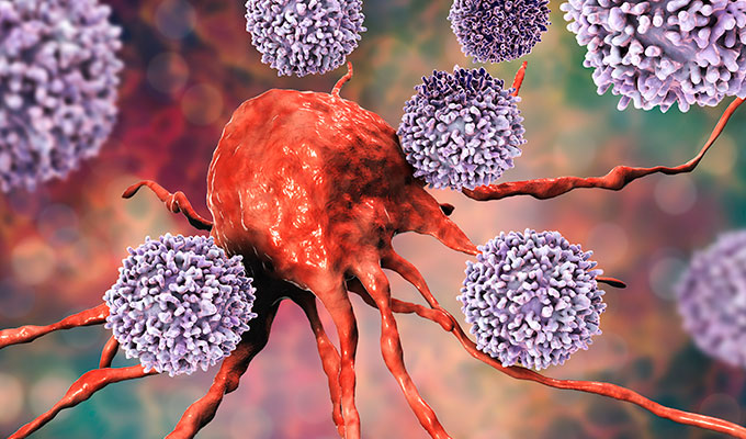
Finding new biomarkers for diagnosis, prognosis or prediction is a hot area in clinical & translational research. Three recent publications are a good example of this.
Li et al. performed a profiling study at the secretome level to differentiate between breast cancer patients and healthy controls, using an antibody array that allows to screen for 274 human secretome biomarkers (including cytokines, chemokines, ECM molecules, growth factors, etc). They found that 19 cytokine levels were significantly different in breast cancer patients. Out of these factors, IL-8, MIP-1 alpha, MIP-1 beta, MMP-8, resistin, FLRG and BCAM were higher in breast cancer patients, while LAP and TSH-β levels were lower.
All markers were clinically validated in breast cancer patients by ELISA, following the usual protocol of performing a profiling array on a small subset of samples (or conditions), and then validating the markers of interest (and only these) in a larger cohort.

For metastatic colorectal cancer (CRC), Hu et al. took a similar approach, this time using an antibody array allowing to study 1,000 secretome biomarkers. Growth and invasion of CRC cells in the liver depend on the microenvironment, so they studied cell culture supernatants from human hepatic sinusoidal endothelial cells (HHSECs). They found that some biomarkers secreted by HHSECs (but not by CRC cells) promoted migration and epithelial-mesenchymal transition, as well as facilitating proliferation and apoptotic resistance of CRC cells. They found that IGFBP-7, Smad 4, SPARC, thrombospondin (TSP), Ras and especially MIF, are mediators whose expression levels were significantly higher in HHSECs than in HUVECs and CRC cells.
Mullen and co-workers used the same antibody array as Hu et al., but with a slightly different approach. In this case, they developed an antibody array allowing to identify novel glutathionylated proteins in the secretome to analyse the redox status of individual proteins. Basically, they incorporated biotinylated glutathione (GSH) into proteins, and then bound them to the above mentioned antibody array. This way, they identified 38 secreted and 55 intracellular glutathionylated proteins. They conclude that comparison of the redox array with conventional proteomic methods provides a higher sensitivity, and it can be performed using more than 100-fold less protein than is required for methods based on mass spectrometry.
Would you like to apply these biomarker profiling tools in your project? Contact your local Biomarker specialists for advice on the best solution!
 Interested in learning more about tools like this?
Subscribe to thematic newsletters on your favourite research topics.