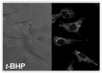
Mechanisms leading to cellulite formation is complex. It involves lipid regulatory pathways and proinflammatory cross-talk that represent promising molecular targets in cosmetology. This post introduces a clever in vitro adipocyte-based assay targeting adipocytokines to better determine the anti cellulite effects of cosmetics compounds.
Among all the molecular pathways followed by skin care professionals, adipolysis / adipogenesis balance in adipocyte tissues is undoubtely one the most attractive ones. Lipolysis induction transforms Triglycerides (TG) contained in adipocytes into Free Fatty Acid (FFA) and Gycerol. It decreases adipocyte size and reduces cellulite phenomenon. Research programs in cosmetology track and record any data related to active compound able to induce direct lipolysis.
In parallel, PPARγ is another key target in cellulite modulation. PPARγ is involved in adipocytes differentiation. Cosmetics strive to identify PPARγ inhibitors to also reduce the number of mature adipocyte tissues stocking fats and located under the skin.
A new molecular target in cosmetology: adipocytokines

Adipocytokines (adipokines) induce inflammatory reactions in adipocyte tissues. Cellulite and obesity stimulate adipocytes into an inflammatory status through the release of proinflammatory adipokines and cytokines. This proinflammatory environment (with MCP-1, IL-6, Leptin, PAI-1, TNFα…) also impairs vascular permeability leading to edema (e.g. heavy-feeling legs) and various other pathophysiological disorders. (1)
Those proinflammatory players have gradually become molecular targets in cosmetology programs. But how to easily and reliably assess proinflammatory effects of cosmetics compounds?
Anti cellulite in vitro evaluation of cosmetics compounds
tebu-bio laboratories have recently developed a unique animal-free method to assess anti cellulite effects of compounds of interest.
This experimental model mimicks in vitro adipokine-related inflammatory process and is available as a fee-for-service. Its design involves 3 main elements:
1- A unique Adipocyte biobank
Having access to a large choice of fully characterized primary cells is the key element. tebu-bio have access to a braod range of Adipocytes from human donors with different Body Mass Index (BMI) ranging from normal, overweight or obese profile purified from multiple body zones (omental and subcutaneous).

This increases the interest of this model by offering more options in the choice of body zone as well as BMI status analyzed in vitro.
2- Appropriate cell culture conditions
Once amplified and plated, selected primary adipocytes can be cultivated under hypoxia or normoxia conditions.
Hypoxia conditions can be reproduced in vitro via a dedicated Hypoxia workstation which allows 100% controlled oxygen level conditions.
At that stage, adipocytes are incubated with the compounds of interest and/or reference compounds.
3- Proinflammatory biomarker profiling & quantification

By using the protein array technology, proinflammatory adipokines can be easily quantified directly from adipocyte supernatants.
The biomarkers thus detected can rapidly characterize the active compound’s efficiency on proinflammatory players according to the chosen adipocyte model. (2)
Adipocytokine protein arrays allows to screen 62 or 182 different biomarkers in a single experiment, offering a unique asset to identify active compounds effects on a complete panel of targeted biomarkers.
Compared to traditional western blot/ELISA approaches, researchers in cosmetology can have access to a far broader range of data without jeopardizing budgets and project development.
Interestingly, Mennesson E. et al. have developped a smart approach to perform both adipokine protein and miRNA profilings in in vitro adipocyte hypoxic models mimicking the physiological state of adipose tissues. (2)
References
- Obesity and its metabolic complications: The role of adipokines and the relationship between obesity, inflammation, insulin resistance, dyslipidemia and nonalcoholoc fatty liver disease, Un Ju Jung and Myung-Sook Choi, Int. J. Mol. Sci. 2014, 15, 6184-6223
- miRNA and protein biomarkers in hypoxia for obesity studies, Éric Mennesson, Isabelle Fixe, Alexandra Foucher, Flavien Carpentier, Nadia Normand (Presented at the 4th Munich Biomarker conference – November 25th-26th, 2014)
Over the last years, various in vitro assays for evaluating the effects of compounds on molecular pathways and for quantifying skin-related biomarkers (Collagen assays, Elastin measurements, Hyaluronic acid quantifications…) have been made available. To further discuss your needs regarding these experimental platforms in cosmetology, get in touch with me.
[contact-form to=’frederic.dubor@tebu-bio.com’ subject=’Adipocytokines in cosmetology’][contact-field label=’Name’ type=’name’ required=’1’/][contact-field label=’Email’ type=’email’ required=’1’/][contact-field label=’Comment’ type=’textarea’ required=’1’/][/contact-form]