As they say, a good picture is worth a thousand words. That’s to say that I’ll have to keep the introduction short in this post (!) to let you know about this interesting map from Boster Bio summarizing the allergic response pathway. I’ll take this opportunity to mention a few related immunoassays that may help you deciphering this pathway in your own study!
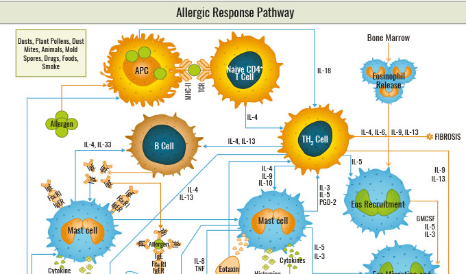
Allergic Response pathway
The body’s immune system may react with a hypersensitive response towards innocuous substances called allergens. Common allergens include dust mites, pollen, mould, food, and animal dander, which can trigger allergic disorders such as allergic asthma, hay fever, allergic rhinitis, anaphylaxis, allergic conjunctivitis, and contact dermatitis. When a susceptible individual is newly exposed to an allergen, the immune system undergoes allergic sensitization. The antigen-presenting cell (APC) complexed with MHC II captures, processes, and displays the allergen to CD4+ T cells to initiate an immune response with Th2 cells. The Th2 cells release cytokines like IL-4 and activate B cells to produce the antibody immunoglobulin E (IgE). Secreted IgE bind to Fc receptors called FcεRI on mast cell and basophil surfaces, which completes the process of sensitization.
When the immune system encounters the same allergen, the allergen binds to IgE and causes cross-linking of IgE and FcεRI. This interaction activates mast cells or basophils to degranulate inflammatory chemical mediators such as histamine, cytokines, and leukotrienes. These mediators invoke vasodilation, mucus secretion, nerve stimulation, and smooth muscle contraction. After the early or acute allergic response, recruitment of other leukocytes (e.g. eosinophils) to the initial site of allergen challenge may induce late allergic responses.

Download the Allergic Response pathway map here.
Which products to decipher the allergic response pathway?
- BosterBio PicoKine ELISAs: Boster takes great measures to ensure product quality and to help you generate comprehensive data upfront. Boster antibodies are validated using WB, IHC, and flow cytometry against a panel of over 250 tissues and un-transfected cell lines to ensure high affinity and crystal-clear IHC stains. In addition, these antibodies are also tested in a quantitative fashion on known quantities of recombinant proteins so that you know what to expect (e.g. if there is 1ng, 2.5ng or 5ng of the target protein in the sample).
- Raybiotech Human Cytokine Array G3: Profiles 42 Human Cytokines in a single experiment: ENA-78, G-CSF, GM-CSF, GRO, GRO alpha, I-309, IL-1 alpha, IL-1 beta, IL-2, IL-3, IL-4, IL-5, IL-6, IL-7, IL-8, IL-10, IL-12 p40/p70, IL-13, IL-15, IFN-gamma, MCP-1, MCP-2, MCP-3, M-CSF, MDC, MIG, MIP-1 delta, RANTES, SCF, SDF-1, TARC, TGF beta 1, TNF alpha, TNF beta, EGF, IGF-1, Angiogenin, Oncostatin M, Thrombopoietin, VEGF-A, PDGF-BB, Leptin
- Raybiotech Human Th1/Th2 Array Q1: Quantifies 10 Human with one array: GM-CSF, IFN-gamma, IL-10, IL-13, IL-2, IL-4, IL-5, IL-6, IL-8 (CXCL8), TNF alpha.
These arrays are suitable for all liquid sample types: serum, Plasma, Cell Culture Supernatants, Cell Lysates, Tissue Lysates and many other Body Fluids. They can be easily outsourced at tebu-bio’s laboratories based in Europe, allowing safe shipment of your samples with no risk of customs delays. tebu-bio is a certified platform for these array services, ensuring quality data and expert support by your designated project manager.
Many other solutions are at hand, including standard ELISA. Just contact me if you need help in choosing the right immunoassay for your research, I’ll be happy to suggest the best solutions!