
When compared to 2D conventional cell culture systems, experimental data obtained with 3D cell culture models have been shown in many instances to be more biologically relevant, closer to clinical conditions (eg. predictive assays/preclinical therapeutic testing).
In this context, tebu-bio laboratories have developed a spheroid platform with a selection of downstream analysis applications such as live cell imaging, microfluidic and physioxia culture conditions or downstream multiplex biomarker quantification, among others.
If you need more physiologically relevant models for your study, but you don’t have access to the technologies or haven’t the time to spend on setting up spheroid cultures in your own laboratories, read on to see what tebu-bio laboratories can generate for you in terms of data.
In this example, tebu-bio’s scientists generated spheroids from cancer cell lines (HT29, LS174T and HCT116), cultured them under different oxygen level conditions (21% and 3%) and compared cancer related biomarker expression to check influence of physioxia, using Raybiotech MMPs Quantibody array QAH-MMP-1.
Generation of the spheroids was previously illustrated in this poster presented at the AACR2018 meeting (click to download). Spheroids were cultured for 7 days before incubation with either DMSO or Mitomycin C. Culture supernatants were harvested after 2 days and incubated on Quantibody following RayBiotech protocol. TIMP1 was detected in the medium surrounding the spheroids above control levels. The differential expression of the protein relative to the oxygen concentrations demonstrated the significant influence of O2 % on protein expression levels.

In the same poster, comparison data obtained from patient derived tissue sample versus healthy control quantified with the same MMP Quantibody array were shown. Dissociated cells from colorectal cancer tissue were cultured on low adherence plates and supernatant analysed for the presence of MMPs after 7 days culture. MMPs, known to have an impact on cell migration and to be upregulated in cancer, were detected, as well as TIMP family proteins, at higher levels in the tumor tissue as expected.

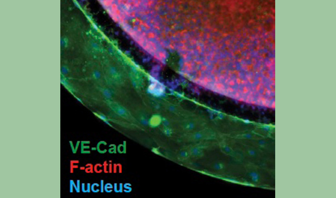
The data shown above are aimed at showing examples of what can be done on this spheroid platform in terms of downstream analysis. More Live cell imaging, microfluidics and transcriptomics data are also illustrated in the poster. tebu-bio project managers can adapt to your own experimental design.
Contact me if you would like to know more or wish to discuss your own project. I’ll be pleased to have a chat with you, or put you in direct contact with my colleague Dora Sabino, our project manager for 3D cell culture studies, first author of the poster cited above.


