2014 Nobel Prize in Chemistry for "nanoscopy"

Atto-550 labeling in Immuno-Fluorescence tebu-bio

The Royal Swedish Academy of Sciences has awarded the 2014 Nobel Prize in Chemistry to Eric Betzig (Janelia Farm Research Campus, USA), Stefan W. Hell (MPI Biophysical Chemistry & DKFZ, GE) and William E. Moerner (Stanford University, USA) “for the development of super-resolved fluorescence microscopy”.

2 separate principles rewarded

  • STimulated Emission Depletion (STED) microscopy (Stefan Hell)
  • Single-molecule microscopy (Eric Betzig & William Moerner)

Single-molecule microscopy

STED-microscopy principle

These fluorescent-based methods allow microscopy to enter into the “nanoworld” (nanoscopy instead of microscopy)  and to give Life scientists hyper-resolutive images.

Source

The Nobel Prize in Chemistry 2014 – Eric Betzig, Stefan Hell, William Moerner. Nobelprize.org

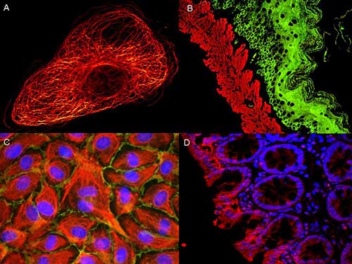
Galery of images obtained with the STED microscopy

Evaluation of AKT activation and migration using STED nanoscopy.
Evaluation of AKT activation using STED nanoscopy and mouse anti-AKT pS473 (p/n 200–301–268, Rockland) and ATTO 647N conjugated anti-Mouse IgG. Source: Leica Microsystems.
Dual color STED microscopy
Courtesy of Leica Microsystems / Rockland Immunochemicals.

605-451-013-Goat-IgG-Antibody-ATTO-425-Conjugated-Rockland-tebu-bio-IF Rockland tebu-bio
ATTO 425 conjugated anti-Mouse IgG was used to demonstrate 2 color STED immunofluorescence microscopy. Methanol fixed A431 cells probed with an anti-tubulin detected with ATTO 425 conjugated anti-MOUSE IgG (610-151-121) secondary antibody (colored RED). In green, anti-HDAC-1 [RABBIT] (p/n 600-401-879) detected with DyLight™488 conjugated anti-RABBIT IgG secondary antibody. Source: Myriam Gastard – Leica Microsystems & Rockland Immunochemicals.

Other high resolutive images obtained with ATTO dyes

Atto-550 labeling in Immuno-Fluorescence tebu-bio
A/ Tubulin in PtK2- male Rat Kangaroo Kidney Epithelial Cells was detected using ATTO 532 labeled secondary antibody. B/ Muscle alpha-actin stained with a mouse primary antibody and ATTO 488 anti-mouse IgG (green) – Cytokeratin stained with polyclonal rabbit anti-cytokeratin and ATTO 647N anti-rabbit IgG (red). C/ HUVEC (Human umbilical vein endothelial cells were stained with anti- Vimentin-ATTO 532 (green), anti-E-Cadherin-ATTO 655 (red) and DAPI (blue). D/ Rat colon sections stained with Anti-Aquaporin 3-ATTO 594 antibody. Hoechst 33342 (blue) is used as counterstain. Source: Dr. Reichwein ATTO-TEC GmbH – Rockland Immunochemicals

views

Leave a Reply

Your email address will not be published.

Subscribe to our newsletter