Immune checkpoint molecules play an important role in T cell functionality after TCR/MHC signalling. Blockade of two B7/CD28 family checkpoint molecules, CTLA-4 and PD-1, have already demonstrated excellent efficacy in increasing T cell responses to a variety of tumours. Identification of novel target and new checkpoint blockade remains a key element in Drug Discovery (See “Drug discovery: Immunotherapy checkpoint research for new Cancer treatments“). In this post, let’s take a look at the “Quantibody® Human Immune Checkpoint Molecule Array 1” for the detection of 10 Human Immune Checkpoint biomarkers.
Approved anti-CTLA-4 monoclonal antibody (Ipilimumab, 2011) and two anti-PD-1 monoclonal antibodies (Pembrolizumab 2014, and Nivolumab 2014) are good examples of new immune Checkpoint-based therapeutic strategies for treating advanced melanoma. Nevertheless, other immune checkpoint molecules have been increasingly considered as new targets for cancer immunotherapy. To identify novel target and new checkpoint blockades, RayBiotech has recently launched the first human Immune Checkpoint Molecule array for detection of the 10 main B7/CD28 family checkpoint molecules: B7-1/CD80, B7-2/CD86, B7-H1/PD-L1, B7-H2/ICOSL, B7-H3, CD28, CTLA-4, ICOS, PD-1, and PD-L2/B7-DC.
The traditional method for cytokine detection and quantification is via ELISA tests for a single target protein (a.k.a. biomarker). Usually, the overall procedure is time consuming and requires a relatively high volume of sample. Thus, conservation of precious small sample quantities becomes a challenging task.

Principle of Quantibody® arrays
Leader in the field of planar protein and antibody arrays, RayBiotech have designed Quantibody® arrays for multiplexed sandwich ELISA-based quantitative array platforms. They allow to accurately determine the concentration of multiple target proteins (eg. cytokines, Immune-Checkpoints…) simultaneously by combining the advantages of the high detection sensitivity & specificity of ELISA and the high throughput of arrays.
Like a traditional sandwich-based ELISA, Quantibody uses a pair of cytokine specific antibodies for detection. A capture antibody is first bound to the glass surface. After incubation with the sample, the target cytokine is trapped on the solid surface. A second biotin-labelled detection antibody is then added, which can recognize a different epitope of the target protein (see figure on the left).
The target protein-antibody-biotin complex can then be visualized through the addition of the streptavidin-conjugated Cy3 equivalent dye, using a laser scanner.
Unlike the traditional ELISA, Quantibody products use an array format. By arraying multiple target proteins specific capture antibodies onto a glass support, quantitative, multiplex detection of proteins of interest in one experiment is made possible.
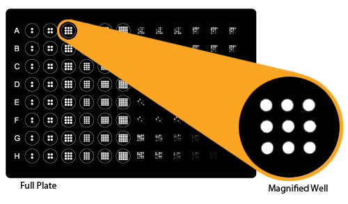
In detail, one standard glass slide is divided into 16 wells of identical cytokine antibody arrays. Each antibody, together with the positive controls is arrayed in quadruplicate. The slide comes with a 16-well removable gasket which allows for the processing of 16 samples on one slide.
Four slides can be nested into a tray, which matches a standard microplate footprint and allows for automated robotic high throughput process of 64 arrays simultaneously.
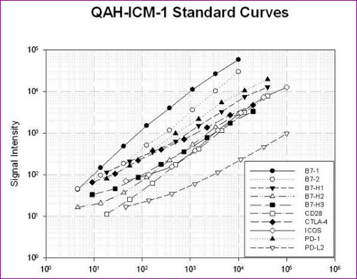
For target protein quantification (ex. cytokines), the array specific cytokine standards, whose concentration has been predetermined, are provided to generate a standard curve for each cytokine.
In a real experiment, standard target proteins and samples will be assayed in each array simultaneously through a sandwich ELISA procedure. By comparing signals from unknown samples to the standard curve, the concentration of the protein of interest in the samples will be determined.

Cytokine Quantibody® array kits have been confirmed to have similar detection sensitivity as traditional ELISA. The current high density Quantibody kits allow scientists to quantitatively determine the concentration of 1000 human, 200 mouse, and 67 rat cytokines in a single experiment.
▶︎ Browse all RayBio’s Cytokine Quantibody Arrays and order them from tebu-bio.
About the new Human Immune Checkpoint Molecule Array
For the new Human Immune Checkpoint Molecule Array 1, 10 human target proteins (CD274, CD276, CD28, CD80, CD86, CTLA4, ICOS, ICOSLG, PDCD1, PDCD1LG2) can be detected and quantified simultaneously in serum/plasma, supernatants, cell & tissue Lysates and many other biological fluids.
Available as 1, 2, or 4 slide kit (16 sub-arrays per slide), the Quantibody Human Immune Checkpoint Molecule Arrays require low sample volume (50 µL or less) and allow to get accurate results within the same day (6-hour processing time).
- Reference: QAH-ICM-1-1: Human Immune Checkpoint Molecule Array 1 (1 array)
- Reference: QAH-ICM-1-2: Human Immune Checkpoint Molecule Array 1 (2 arrays)
- Reference: QAH-ICM-1-4: Human Immune Checkpoint Molecule Array 1 (4 arrays)

Immune checkpoint targets & assays (BPS Bioscience – tebu-bio). Download BPS poster – Immuno-Oncology Summit – Boston 2017. - Reference: QAH-ICM-1-SW: Q-Analyzer Software
Related products related to research about Immuno-therapies & Immune checkpoints:
- Human Gastric Cancer Biomarker Array Q1 (cat. nr QAH-GCB-1) – Detects 5 human gastric cancer biomarker proteins (PGC, PGA5, PGA4, PGA3, CEACAM5, CA (72-4), CA (19-9)).
- Human Cancer Biomarker Array 1 (cat. nr QAH-CAN-1) – Detects 18 human cancer biomarker proteins (Alpha-fetoprotein, CA125, CA15-3, CA19-9, CA72-4, CEA, Cytokeratin 19 (KRT19), GOLM1 (GP73), hCG beta, HE4 (WFDC2), NSE, Pepsinogen 1, Pepsinogen 2, ProGRP, PSA-free, PSA-total, SERPINB3 (SCCA1), Thyroglobulin).
- Biochemical and Cell based assays tools for screening and validating new molecules against immune checkpoint pathways. More specifically, BPS Bioscience have developed Luciferase cell based reporter assays to identify new drugs candidate over several human Immune checkpoints and enzymatic pathways:
-
- PD-1:PD-L1 – Programmed Cell Death 1
- TIGIT:CD155 – T-Cell Immunoreceptor with Ig and ITIM domains
- GITR:GITRL – Glucocorticoid-induced TNFR Related protein
- OX40:OX40L – TNFRSF4, CD134
- CD40:CD40L – Cluster of Differentiation 40, TNFRSF5, Bp50
- LAG3:MHCII – Lymphocyte-Activation Gene 3

AIM Biotech’s chips for assessing tumor immune response ex vivo tebu-bio (Application notes available here) To get more insights on these Immune Checkpoint cell based assays, the poster “Methods for Drug Discovery Through Biochemical and Cell Based Assays for Immunotherapy Checkpoint Activators and Inhibitors”presented by BPS Bioscience at the Immuno-Oncology Summit in Boston this year (2017)
- Download the poster here
▶︎ Browse all BPS Bioscience’s reporter cell lines and order them from tebu-bio.
- Innovative cell culture devices for more predictive 3D models for assessing tumor immune response with Aim Biotech‘s chips ex vivo
Download the Application Note here
Outsource your biomarker discovery
 Your protein multiplex quantification can be performed by tebu-bio’s laboratories.
Interestingly, these Quantibody arrays are regularly used by our own scientists. Therefore we can perform these multiplex quantifications in our laboratories. Just send your samples to us, and get the data you need back in a few days.
Interested? Don’t hesitate to contact us for further information!