
Recently, a we published a series of posts focusing on research tools aiming at studying actin through biochemical assays and in fixed and living cells (e.g. Actin visualization, Actin binding, Actin polymerization, and measurement of the G:F actin ratio in cells).
Today, this is the first post on how to vizualize tubulin in cells. This is the first publication of a series, that will be dedicated to tubulin visualization, binding and polymerization as well as measurement of the tubulin vs. microtubule ratio.

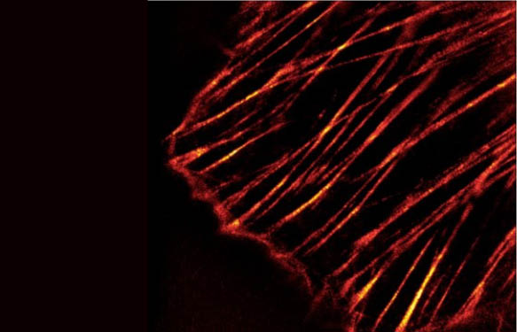
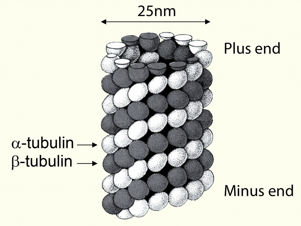
Tubulin represents one of the major cytoskeleton structures. It plays an important role in cell structure, intracellular transport, and mitosis. In eukaryotic cells, microtubules are assembled from dimers of α-tubulin and β-tubulin which form a filamentous cylinder. While β-tubulin is exposed on the (+) end, α-tubulin is exposed on the (-) end. As filamentous F actin, microtubules can grow on both (+) and (-) ends, although the growth at the (+) end is much more rapid.
Tubulin visualization and detection by immunocytochemistry
Tubulin detection can be made through immuno-assays such as IP / Western Blots, ELISA, and Immunocytochemistry. One needs 100% guaranteed primary antibodies to do so. An overview of such research antibodies from specialized sources can be found here.
Of particlar interest, Cytoskeleton’s Anti-alpha/beta tubulin has been positively tested in the all these applications and belongs to the most popular antibody.
Tubulin / microtubule fluorescent dye for live cell imaging

Spirochrome’s SiR-Tubulin fluorescent dye is a tubulin-specific cell permeable compound. It allows microtubule vizualization without the need to transfect cells with vectors carrying the information for fluorescently labeled Tubulin or binding proteins.
A few experimental results obtained with Spirochrome’s SiR stain can be seen in this previous post User experience of SiR-Actin and SiR-Tubulin Live Cell Imaging.
Interested in visualizing Tubulin?
Get in touch by leaving your questions or comments below!