
Page 2 - Scientific Library
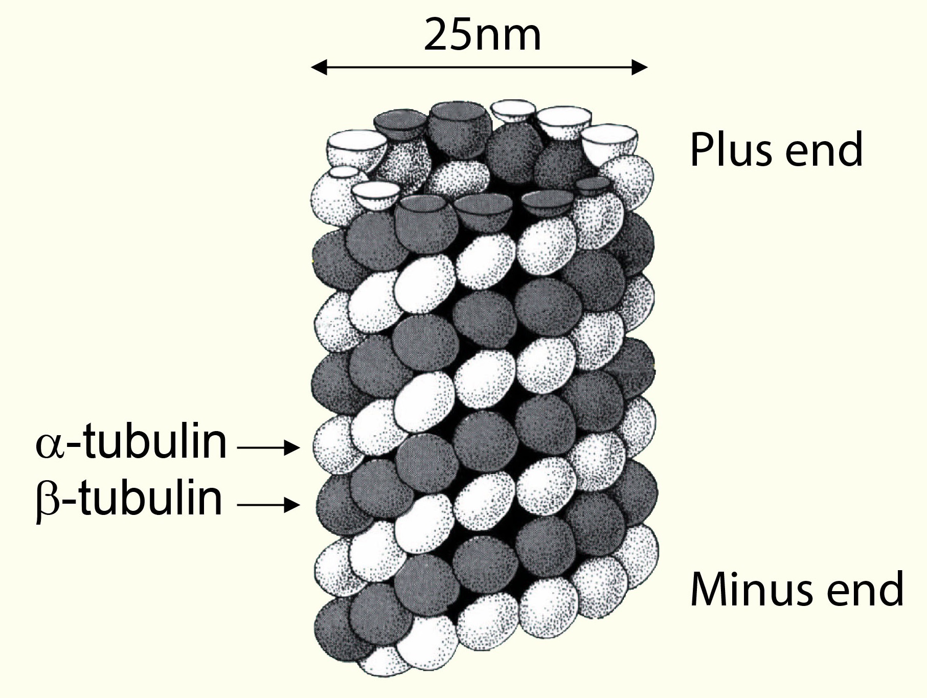
Versatile measurement of protein binding to microtubules
You suspect your protein binds to microtubules? That it might stabilize or destabilize these filamentous structures? Then this post is here to help you to find a meaningful assay to validate your assumption.
How sample preparation can affect your biomarker studies (II)
In a previous post, we looked at the importance of sample preparation methods depending on the biomarker being studied (e.g. MMPs, cytokines). Today, let's focus on cell / tissue lysates.
Tip #1
Tubulin polymerisation measurement - made easy!
Recently, I issued a post about a method which allows measuring microtuble binding capabilities of proteins of interest. Today, I invite you to look at methods for
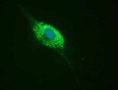
Tubulin:Microtubule ratio measured in cells
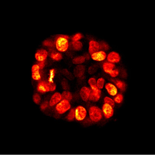
Live cell imaging of Actin, Tubulin & DNA - SLAS conference
In the autumn of 2014, we presented new stains to conduct live cell imaging of Actin and Tubulin, which I covered in my post 2 new Actin and Tubulin live-cell imaging stains – without transfection!
Post-Translational Modifications (PTM) of Tubulin
In this post, I'd like to take a look at the current understanding of tubulin PTMs, that include tyrosination/detyrosination, Δ2-tubulin formation, acetylation, phosphorylation, ubiquitination, glutamylation,
SiR fluorogenic probes: multicolour live-cell Imaging of Actin, Tubulin, DNA, and Lysosomes
3 functional assays to investigate actin dynamics
Today, I'd like to give you an overview about methods in actin research with validated R&D products and kits which will allow you to measure binding to actin and effects on the polymerisation dynamics
Fluorescence microscopy: how to improve your Live Cell Imaging
For more than 2 years now, the Silicon Rhodamine-like (SiR) technology has allowed the live cell imaging field with fluorescence microscopy to evolve significantly.
Fluorescent SiR-probes have appeared