

Hyaluronic acid (Hyaluronan) belongs to the group of glycosaminoglycans (GAGs), but unlike other GAGs like Heparan sulfate, Chondroitin sulfate or Keratan sulfate, it cannot be found as a proteoglycan. It represents a non-sulfated polysaccharide consisting of alternating residues of D-glucuronic acid and N-acetylglucosamine (Fig 1). In the human umbilical cord and synovial fluid, the size of HA is reported to be about 3,000,000 Da (a flexible chain of 4000 disaccharide units).
HA is one of the major components of the extracellular matrix and it absorbs high amounts of water thus giving tissues the ability to resist compression. Furthermore, Hyaluronan contributes significantly to cell proliferation and cell migration. As HA levels often correlate with malignancy and poor prognosis of certain cancers, it can be used as a tumor marker. As Hyaluronan plays a role in skin healing, it’s a widely used ingredient of skin-care products.
In recent posts, I presented versatile, colorimetric methods to measure the other main components of the extracellular matrix, such as

Purple-Jelley™ – the easy-to-run assay to measure Hyaluronic acid
Today I will concentrate on a new assay recently released by Biocolor which allows the measurement and quantification of Hyaluronic acid from tissues. The assay relies on the affinity of the Purple dye (3,3’-Diethyl-9-methyl-4,5,4’,5’-dibenzothiacarbo-cyanine bromide, Fig 2) to Hyaluronic acid. To avoid cross reactivity of the dye, removal of tissue protein before assay is essential in order to isolate the HA from potentially dye binding proteins. Moreover sulfated glycosaminoglycans (sGAG) need to be removed before measuring the isolated HA. Methods for protein and sGAG removal are specified in the Purple-Jelley™ assay manual. Finally the readout of the assay can be done at 655 nm using a microplate spectrophotometer.
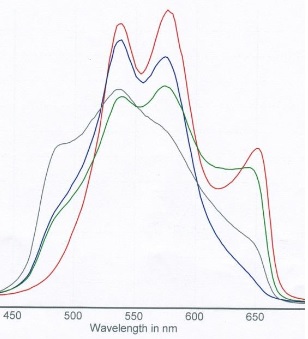
A read-out of Hyaluronic acid alone, chondroitin 4-sulfate alone and mixture of both GAGs is shown in Fig 3, which shows the need to remove sulfated GAGs from samples to get a clearly separated HA peak at 655 nm. A typical standard curve with isolated Hyaluronic acid (provided in the kit) is shown in Fig 4.


Interested in measuring Hyaluronic acid or any other of the matrix components mentioned above? Contact me through the form below for further information.
 Interested in learning more about tools like this?
Subscribe to thematic newsletters on your favourite research topics.


One Response
Hi, I would like to test this dye for measuring and quantifying HA samples.