As is well known, the actin cytoskeleton is a highly dynamic structure, involved in a large number of cellular processes, including muscle contraction, lamellopodial extrusion, cell locomotion, cytokinesis, intracellular transport and many more. Under the tight regulation of more than 50 actin binding proteins (ABPs) and in response to a wide variety of internal and external stimuli, the morphology of the actin cytoskeleton changes rapidly.
![]() For more than 20 years now, Cytoskeleton Inc. has provided a large selection of robust and user friendly solutions (pure and active actin proteins/Biochemical assays and actin staining probes..) which have helped many researchers to solve fundamental questions in cell signalling, cytoskeleton and actin research fields.
For more than 20 years now, Cytoskeleton Inc. has provided a large selection of robust and user friendly solutions (pure and active actin proteins/Biochemical assays and actin staining probes..) which have helped many researchers to solve fundamental questions in cell signalling, cytoskeleton and actin research fields.

Actin is a 43 kDa protein that is very highly conserved between species. There are three main actin isotypes (α, β and γ), which show >90% amino-acid (aa) homology between isotypes and >98% homology within members of a particular isotypic group. As shown in Fig. 1, Globular-actin (G-actin) readily polymerizes under physiological conditions to form Filamentous-actin (F-actin / double-helical filament) with the concomitant hydrolysis of ATP.
By providing easy access to some of the most powerful in vivo and in vitro products employed in the field, tebu-bio helps you in your daily work to better understand actin behaviour and interactions. Let’s take a look at a small selection of such tools.
Actin Proteins
Cytoskeleton offers unmatched activity and purity in the most comprehensive actin portfolio.

- Choice of >95% or >99% pure
- Labeled or unlabeled proteins
- Cytoplasmic, and cardiac, skeletal and smooth muscle sources
- Bulk quantities available
As presented in Fig. 2 for the Actin purified from skeletal muscle (AKL99 >99% pure and AKL95 >95% pure), purity is determined by scanning densitometry of Coomassie Blue stained protein on a 12% polyacrylamide gel. The ability of this protein to efficiently polymerize into filaments (F-actin) in vitro (Biological activity) has been determined by a spin down assay (separate F-Actin from unpolymerized components). Stringent quality control ensures that AKL99 produces >90% F-actin and AKL95 produces >80% F-actin in this assay.
Biochemical kits

Cytoskeleton’s Biochem Kits™ are comprehensive kits for assaying different aspects of cytoskeletal biochemistry and signal transduction:
- Measuring the effects of proteins and drugs on actin polymerization/depolymerization.
- Fluorescent Actin Polymerization Biochem Kit (Functional assay) – Fig.3
- Discover and characterize actin binding proteins (ABPs).
- BK001 – muscle actin binding proteins (Functional assay)
- BK013 – non-muscle actin binding proteins (Functional assay)
- Quantitate G-actin (monomer) vs F-actin (polymer) in cell or tissue lysate.
- G-actin / F-actin in vivo assay kit (Cell-based assay)
- Minimize set-up time from months to days.
- …..
These kits usually form the basis of a figure in a publication. They come with all the reagents needed for your assay as well as detailed instructions on how to use them, so you’ll be ready to do your assays as soon as you receive the kits! More technical information on these assays is available in our previous post.
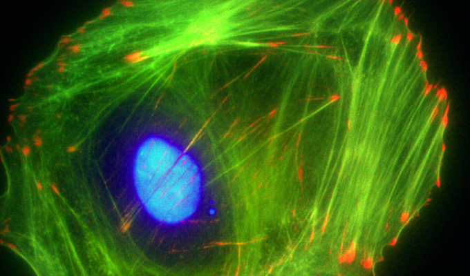
Fluorescent Actin probes
Actin staining can be very useful for determining the structure and function of the cytoskeleton in living and fixed cells. The actin cytoskeleton is a very dynamic and labile structure in the living cell, but it can be fixed with paraformaldehyde prior to probing or staining for actin structures. For fixed cell staining, Cytoskeleton have developed the Acti-stain™ line of fluorescent phalloidin probes which are exceptionally bright and stable (Fig. 4), allowing you to:

- Stain F-actin in fixed cells
- Stabilize actin filaments in vitro
- Visualize actin filaments in vitro
Other then being exceptionally bright, these probes are photostable and also compatible with popular filter sets such as FITC, TRITC and Cy5.
- Acti-stain™ 488 phalloidin-PHDG1-A
- Acti-stain™ 555 phalloidin-PHDH1-A
- Acti-stain™ 670 phalloidin-PHDN1-A
The combination of in-house manufacturing, stringent quality control and convenient packaging provides great value.
You might be also interested in labeling actin and cytoskeleton on living cells. In this case, you might like to read our posts on the fluorescent probe available for live imaging of actin.
And even more actin proteins and kits…
This is only a summary of a small selection of all the Cytoskeleton Inc. products available for your cytoskeleton and actin studies. To go further in-depth and to help you to find the right (and best!) tools among all the available solutions, take a few minutes to browse our Actin Product Guide.
Of course, don’t hesitate to contact your local tebu-bio office, or to leave your questions and comments below!