Introduction
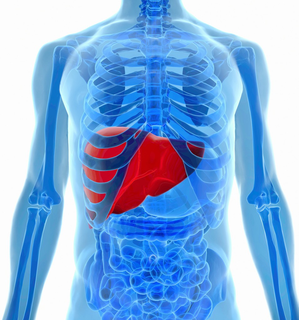
Hepatic stellate cells (HSC, also known as perisinusoidal cells or Ito cells), are liver-specific mesenchymal cells found in the space of Disse. HSC maintain interactions with sinusoidal endothelial cells and hepatic epithelial cells. The stellate cells are involved in liver physiology and fibrogenesis (formation of scar tissue in response to liver damage).

In a healthy liver, stellate cells are described as being in a quiescent state, and represent 5-8% of the total number of liver cells. (1) Each cell has several long protrusions that extend from the cell body and wrap around the sinusoids. Lipid droplets formed in the cell body store vitamin A, constituting the largest reservoir of vitamin A in the body. (1)
Hepatic inflammation

During hepatic inflammation, triggered by virus infection or immune-mediated mechanisms, hepatic stellate cells receive signals secreted by damaged hepatocytes and immune cells, causing them to transdifferentiate into activated myofibroblast cells (2). Fibrogenesis is initiated by inducing apoptosis of hepatocytes. This has mechanism was already described into a recent post (http://5.196.212.231/2014/04/15/activation-de-activation-pathways-of-hepatic-fibrosis/).
The activation process is fueled by the release of lipid droplets (autophagy). The apoptotic bodies from the damaged hepatocytes can also stimulate Kupffer cells to generate reactive oxygen species and nitric oxide by inducible nitric oxide synthase. The ROS in turn can enhance hepatocyte apoptosis and continue the activation of hepatic stellate cells. As the primary extracellular matrix–producing (ECM-producing) cells in liver, activated stellate cells generate a temporary scar at the site of injury to protect the liver from further damage.
Clinical studies indicate that hepatic fibrosis is reversible (3-5). During the regression of fibrosis, the number of activated hepatic stellate cells is greatly reduced by the induction of cellular senescence and apoptosis, or by the return to the quiescent state (4-7).
Because of their pivotal roles in liver repair and disease pathogenesis, hepatic stellate cells have been a major focus of liver research. Therefore sourcing of the adapted cellular model is crucial before starting new experiments.
Cellular models available at tebu-bio
- Cryopreserved human hepatic stellate cells
- Cryopreserved human liver myofibroblasts
- Cryopreserved human Kupffer cells
- Cryopreserved & freshly isolated human hepatocytes
- Freshly isolated non-parenchymental human hepatic cells – with or without matched hepatocytes and/or PBMCs (please contact)
Please contact me to source your cellular models for your next studies.
[contact-form to=’jean-francois.tetu@tebu-bio.com’ subject=’Liver cells – tebu-bio blog’][contact-field label=’Name’ type=’name’ required=’1’/][contact-field label=’Email’ type=’email’ required=’1’/][contact-field label=’Comment’ type=’textarea’/][/contact-form]