In a previous post, I mentioned that the secretome is becoming a very hot topic in the world of proteome analysis, and even a crucial study subject. Today, I’d like go a step further and shed some light on how to analyse it.
Let’s see what the options are…
First point: How to identify proteomic changes induced by the experimental conditions?
The Classic methods:
Mass Spec identification: This combined approach is widely used since it is supposed to be unbiased: no a priori, and identification of all proteins. Well, in theory…
Due to the huge amount of proteins analyzed and the method (cleavage of roteins into peptides before analysis), the low concentration proteins don’t even appear (usually the maximum sensitivity is close to 1.000 pg/ml).
Moreover, this method has its limitations: cost of the equipment and the individual tests, low throughput…
Western blot: a reference technique! It covers all the limitations of the Mass Spec, but the mandatory selection of antibodies implies deciding upfront which analytes to look at.

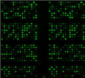
in red, positive control spots give the orientation of the array, and will be used for array normalisation.
Analysis of the results is done by comparing the signal of different conditions (arrays) for each analyte (see blue spots for analyte A, green spots for analyte B).
New alternatives:
Profiling Antibody Arrays:
Antibody arrays: They combine specificity and high sensitivity of Western Blot with multiplex capacity. Antibody arrays show high sensitivity (down to 1pg/ml), are easily accessible (no dedicated device is needed since they are compatible with most micro-array scanners), offer higher throughput at an affordable cost, while allowing the analysis of several hundreds of proteins / assay.
Antibody arrays allow you to study proximal biological fluids for cancer biomarker analysis (low volume and low concentration) requiring use of very sensitive detection methods where Antibody Arrays will do the job perfectly…
Second point:
How does this Antibody Array technique work?
Antibody arrays are membrane or glass-slides spotted with Antibodies specific for secretome proteins, that capture these proteins. The detection is then done in chemiluminescence (membrane arrays) or fluorescence (laser scanner for glass-slides) and allows you to identify the changes occuring between different experimental conditions.
This approach gives a unique opportunity of fishing for new proteins, to become tomorrow’s Biomarkers..
Third and last point:
How to choose between membrane and glass-slides?
 Membrane arrays are read in chemiluminescence, same way as a WB. No heavy equipment is needed.
- Glass-slides arrays require a laser-scanner (that can be found in most labs working with DNA chips) but the samples will be 5-10 times smaller, and the assay is much cheaper per point.
My short-list of suggestions
Of course, as for WB, the use of Antibodies demands a preselection of targets…
Which options do you have currently? Here my favourite ones:
- Broad profiling: 1.000 Human targets in row (Cytokines, Chemokines, adhesion molecules, soluble receptors, matrix proteins…)
- 40 Mouse inflammatory proteins analyzed from 10µl of serum
- Characterization of 43 Human Angiogenic factors, with no equipment at all (membrane array): Angiogenesis Array 1.000
Publications using Antibody arrays to identify new secretome biomarkers
Angela Schulz et al. “Inflammatory cytokines and signaling pathways are associated with survival of primary chronic lymphocytic leukemia cells in vitro: a dominant role of CCL2” (German Cancer Research Center, Department for Molecular Genetics, Heidelberg).
Ramos-Mozo P, Rodriquez C, Pastor-Vargas C, Blanco-Colio LM, Martinez-Gonzalez J, Meilhac O, et al. (2012). “Plasma profiling by a protein array approach identifies IGFBP-1 as a novel biomarker of abdominal aortic aneurysm”. Atheroscler. http://dx.doi.org/10.1016/j.atherosclerosis.2012.01.009 http://www.sciencedirect.com/science/article/pii/S0021915012000135
Chen H, Wang Y, Bai C, Wang X. (2012) “Alterations of plasma inflammatory biomarkers in healthy and chronic obstuctive pulmonary disease patients with or without acute exacerbation”. J Proteom. http://dx.doi.org/10.1016/j.jprot.2012.01.027 http://www.sciencedirect.com/science/article/pii/S187439191200067X
Need more info about these arrays?
Contact our specialists!
[contact-form to=’myantibody@tebu-bio.com’ subject=’Secretome research’][contact-field label=’Name’ type=’name’ required=’1’/][contact-field label=’Email’ type=’email’ required=’1’/][contact-field label=’Comment’ type=’textarea’ required=’1’/][/contact-form]