
Despite its importance in the regulation of many cellular processes, membrane tension is still complicated to measure. And despite valuable insights, the techniques currently available remain complex and present technical limitations. To overcome these drawbacks, Spirochrome have developed Flipper-TR, the first probes for fluorescence microscopy in the field of mechanobiology, that specifically targets the plasma membrane of cells and reports membrane tension through changes in its fluorescence lifetime.
Why study membrane tension in cells?
Cellular and Organelle membranes composed of lipid bilayers are dynamic, fluid and highly deformable structures (4nm thick), allowing cells to change shape and tension and be engaged in physiological functions.
The membrane tension plays a crucial role in cellular processes whether at the whole cell level (migration, spreading, phagocystosis, division), or also at the sub-cellular level (metabolism, mechano-transduction, endocytosis…). As the membrane tension is constantly regulated by the cell, the study of its regulation, and also how it regulates cellular processes over the time and location, is relevant for a better understanding of cellular behavior for both healthy and diseased cells.
Measuring membrane tension
Despite its importance, membrane tension is (or has always been) complicated to measure in cells. The only technique available involves pulling on membrane tubes from the plasma membrane with a bead trapped in an optical tweezer. More than providing valuable insight, this techniques suffers from complexity and several methodological and technical limitations. However, by developing the new Flipper-TR® probe, Spirochrome have taken up the challenge to provide an easy to use tool for a sensitive and real time measurement of membrane tension in living cells(1).
What makes the fluo Flipper-TR probes special?
Flipper-TR® is the first fluorescent membrane tension reporter developed for the field of mechanobiology. The fluorescence lifetime of the probe is strongly dependent on the membrane tension, and through FLIM* (fluorescence lifetime imaging microscopy) a precise measurement of the spatio-temporal distribution of tension in membranes is possible.

Left. Schematic diagram of the mechanism of Flipper-TR
Right. Flipper-TR solution absorbance (dotted orange line) and emission (solid orange line)
How do the Flipper-TR probes work?
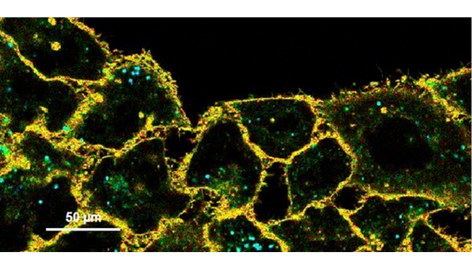
Flipper-TR® specifically targets and inserts into the plasma membrane of cells, and finally is only fluorescent when inserted in a lipid membrane (fig1). Changes in the organization of the lipid bilayer membranes, induces a change of the twist angle and polarization between the two twisted dithienothiophenes of the mechanophore. The Flipper-TR® probe has a broad absorption and emission spectrum, excitation can be commonly performed with a 488nm laser, while emission is collected between 575 and 625nm. Based on its technical characteristics, Flipper-TR® appears as the most advanced member of the Flipper probes family(2,3).

Left: Cells stained with Flipper-TR® before (top) and after (bottom) hyperosmotic shock. Right: The histogram shows the lifetime shift after osmotic shock
Quick and sensitive Flipper-TR
Flipper-TR® finally provides quick and sensitive reports of the changes in the cell’s membrane tension through the modification of its fluorescence lifetime. Changes in the Osmotic environment have a direct impact on the shape, and thereby the membrane tension of the cell (fig 2). During hyperosmotic shock (high osmolarity in culture medium) the volume of the cell decreases leading to a decrease of the membrane tension, and a shift of the fluorescence lifetime (fig. 2- red scale). In Osmotic conditions (fig2- Before OS), we robustly observed a longer lifetime (fig. 2 – grey scale).

Fig. 3: Flipper-TR staining in yeast cells
Flipper-TR® staining in yeast cells. Yeast cells stained with Flipper-TR and treated with either hypo-osmotic shock (left) or hyper-osmotic shock (right)
Flipper-TR® works also on a wide range of organisms including mammalians, bacteria and also yeast(4) cells as shown figure 3. Under Hypo-Osmotic conditions (increase in membrane tension) the fluorescence lifetime shifts to high values, in contrary to hyper-osmotic condition where the lifetime shift to low value (blue) as seen previously on figure 2.
Organelle specific probes
Using the same innovative and revolutionnary technology as for the Flipper-TR® probe, new organelle-specific mechanosensitive Flipper-TR probes which target ER, mitochondria & lysosomes of live cells have also been developed.
Endoplasmic reticulum Flipper-TR probe
ER Flipper-TR® is a fluorescent probe that specifically targets the endoplasmic reticulum (ER) of cells and reports membrane tension changes through its fluorescence lifetime changes. It contains an ER targeting motive as well as a Flipper fluorophore(1). The probe spontaneously labels the ER of cells and is only fluorescent when inserted in a lipid membrane. It has a broad absorption and emission spectrum, excitation can be commonly performed with a 488nm laser, while emission is collected between 575 and 625nm.

Left: FLIM & intensity images of a HeLa cell labeled with ER Flipper-TR
Right:CLSM images of a COS7 cell labeled with ER Flipper-TR, (green) and ER-Tracker Red, (red) and the merged images
Mitochondria Flipper-TR probe
Mito Flipper-TR® is a fluorescent probe that specifically targets the membrane of mitochondria of cells. The probe spontaneously labels mitochondria of live cells and is only fluorescent when inserted in a lipid membrane. It has a broad absorption and emission spectrum, excitation can be commonly performed with a 488nm laser, while emission is collected between 575 and 625nm.

FLIM images of HeLa cells labeled with Mito Flipper-TR before and after osmotic shock
Lysosome Flipper-TR probe
Lyso Flipper-TR® is a fluorescent probe that specifically targets the membrane of lysosomes of cells. The probe spontaneously labels lysosomes of cells and is only fluorescent when inserted in a lipid membrane. It has a broad absorption and emission spectrum, excitation can be commonly performed with a 488nm laser, while emission is collected between 575 and 625nm.

FLIM & intensity images of HeLa cells labeled with Lyso Flipper-TR
Further than providing solutions to the limitations of current methods, with the Flipper-TR® probe, Spirochrome have opened up a whole new field by giving access to real time membrane tension measurements in live cells.
Where can I order Flipper-TR probes?
Contact your tebu-bio local office to get more insight on this innovative probe, and don’t hesitate to take a look at our other Spirochrome probes, or all our Live Cell Imaging solutions including fluorescence microscopy on this interactive web page.
References:
1) Colom A, et al: A fluorescent membrane tension probe. Nat Chem, 2018, 10:1118–1125 ().
2) Dal Molin M, et al: Fluorescent flippers for mechanosensitive membrane probes. JACS, 2015, 137:568-571.
3) Soleimanpour S, et al: Headgroup engineering in mechanosensitive membrane probes. Chem Commun (Camb) 2016, 52:14450-14453.
4) Riggi M, et al: Decrease in plasma membrane tension triggers PtdIns(4,5)P2 phase separation to inactivate TORC2, Nat Cell Biol, 2018 Sep; 20 (9): 1043–1051
*:FLIM microscopy stands for Fluorescent Lifetime Imaging Microscopy. The importance for membrane tension studies is that prior to Flipper-TR membrane tension measurements were very labor and equipment intensive, but now relatively straightforward adaptation of your current microscope will enable highly sensitive tension measurements. Nowadays it is a very standard technique with equipment available from many suppliers, it is based on recording the time that emission takes after excitation of the fluorophore, which is usually very rapid, on the order of 1-10 nano-seconds. FLIM can also be combined with other high resolution microscopic techniques such as Total Internal Reflection Fluorescence (TIRF) or Stimulated Emission Depletion (STED) microscopy for high spatial resolution. FLIM microscopy requires time resolved light detectors which many scientific microscope vendors have available, for example PicoQuant’s upgrade kit (https://www.picoquant.com/news/item/picoquants-flim-fcs-upgrade-kit-now-supports-zeiss-lsm780-and-leica-sp2). Reference 1 describes more details about the experimental setup for FLIM microscopy. (Flipper-TR is a registered trademark of UNIGEM, Switzerland)


