Live Cell Imaging has transformed the way biologists study cellular functions and processes. The observation of dynamic changes provides more insight into the processes happening in a cell, as compared to imaging studies of fixed cells. Goryo Chemical have developed a broad range of innovative Live Cell Imaging fluorescent probes for various applications. Let’s take a closer look…
Stem Cell Research
Kyoto Probe 1 (KP-1): Specifically label human iPS and ES cells

KP-1 probe is cell permeable and mitochondria localized in the human pluripotent stem cells regardless of mitochondrial membrane potential. After the differentiation of the cells, KP-1 is eliminated from the cells by ABC transporters (which do not work in pluripotent stem cells), and the cells become non-fluorescent (find more details here).
- Able to distinguish human iPS cells and/or human ES cells from differentiated cells
- Usable for flow cytometry or live cell imaging
- Staining while culturing
- λex: 515nm – λem: 529nm
Cancer Research

MAR: for detection of hypoxia (reductase)
- Study of cancer or Ischemia
- Easily available for live cell imaging / flow cytometry
- As sensitive as pimonidazole
- λex: 515nm – λem: 529nm

ProteoGREEN-gGlu: for specific detection of cancer cells
- Cancer cells specific
- High signal-to-noise ratio
- GGT (γ-glutamyltranspeptidase)-activable fluorescent probe
- For fluorescent imaging of cancer cells
- λex: 496nm – λem: 525nm
Reactive Oxygen Species Research
Reactive oxygen species (ROS) play key roles in many intracellular processes and are known to be involved in various diseases such as carcinogenesis, inflammation, and many others. Many species of ROS exist and each one is likely to have a specific role in living cells.
HYDROP: for specific detection of hydrogen peroxide

(find more technical details here in our previous post)
- High specificity to hydrogen peroxide (H2O2)
- Suitable for time-lapse imaging because it photo-bleaches slowly
- λex: 492nm – λem: 518nm
Other ROS specific probes:
- Hydroxyphenyl Fluorescein (HPF) for the detection of hydroxyl radicals (· OH)
- Aminophenyl Fluorescein (APF) and NiSPY-3 for the detection of peroxynitrite (ONOO —)
- HySOx for the detection of hypochlorous acid (HOCl)
- OxyOrange (NEW) for specific detection of hydroxyl radicals (· OH) and hypochlorous acid (HOCl).
Inorganic Biochemistry Research
CopperGREEN: for the specific detection of Cu1+

As described in our previous post the CopperGREEN probe is specially designed for:
- High specificity to copper ion: low reactivity with other trace metal ions and reactive oxygen species (ROS)
- High signal-to-noise ratio: fluorescence increase of over 100 times can be observed upon reaction with copper (I) ion
- λex: 440-490nm – λem: 510nm
FeRhoNox-1: for specific detection of Fe2+

Iron is indispensable for our life and is the most abundant transition metal in our body. Iron acts as oxygen carrier or active center of various enzymes. Although iron is crucial to us, its strong oxidation-reduction activity has a toxic effect on cells, and can even cause cell death if present in excess amounts.
FeRhoNox-1 advantages:
- Specific detection of Fe2+
- Applicable to live cell imaging of labile Fe2+
- Detection of iron (II) ion in Golgi complex
- λex: 540nm – λem: 575nm
Other inorganic compound specific probes:
- CaTM-2™/CaTM-2™ AM for red-fluorescent detection of Ca2+
- CaSiR-1™/CaSiR-1™ AM for near-infrared detection of Ca2+
- ZnAF-2/ZnAF-2 DA for the detection of Zn2+
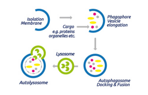
Lysosome – Late Endosome – Secretory Granule Research
AcidiFluor ORANGE: pH probe for acidic organelles

With the Acidifluor™ ORANGE probe, fluorescence increases greatly when under acidic condition. This probe can stain acidic organelles, such as lysosomes, late endosomes and secretory granules.
Advantages of the Acidifluor™ ORANGE probe:
- High signal-to-noise ratio
- Orange color fluorescence and usable for multicolour imaging
- Great photo-stability
- λex: 535nm – λem: 560nm

Β-galactosidase – Senescence Associated-β-Gal Research
GlycoYELLOW-βGal: for detection of β-galactosidase and SA-β-gal
 Usable for Live Cell Imaging
- High sensitivity
- High retention ability in cell
- λex: 525-488nm – λem: 543nm


Other B-gal specific probes:
GlycoGREEN-βGal (NEW) for green fluorescent detection (λex: 497nm – λem: 519nm) of β-galactosidase and SA-β-gal in normal and cancer cells (find more details about this probe here in our previous post)
Super-Resolution Live Cell Imaging
HMSiR: Blinking without high-power laser irradiation

dSTORM (direct stochastic optical reconstruction microscopy) is a super-resolution microscopy technique based on the blinking of fluorescent probes. The HMSiR probe displays spontaneous blinking in light emission, eliminating the need for thiols, oxygen scavengers and high-power laser irradiation from super-resolution imaging.
HMSiR probe benefits:
- Available for super-resolution live cell imaging under physiological conditions without thiols or oxygen scavengers.
- Observation without high-power laser irradiation minimizes damage caused to the cells
- λex: 647nm – λem: 692nm
Find more HMSIR information here.
To conclude…
Any questions about these tools for Live Cell Imaging? Get in touch by leaving your questions or comments below, I’ll be pleased to get back to you.

Looking for other tools for Live Cell Imaging? Try this interactive guide, where you can simply click on the parameter you want to measure to find the associated tools.