Gastrointestinal (GI) primary cells represent a powerful approach for the in vitro study of the physiopathology of this unique tissue. Researchers have now identified intimate interactions between GI cells and their microbia with neurological disorders pointing out the need to access highly characterized sources of GI primary cells to design reliable and more physiologically relevant in vitro cellular models.
Recent discoveries have shown the strong link between the Gastrointestinal tract and neurobiology. A few examples:
- Shift of Gut Microbiome in Early Stage of Parkinson’s disease (PD) – Changes in the gut microbiome occurs long before PD symptoms are observed. Thus, the gut microbiome shifts link intestinal barrier function and immune function in PD patients
- Treating Autism by Gut Microbiota Transplant – A pilot study has revealed that microbiome transplantation eases digestive troubles and improves social skills in children with autism
- Implication of GI-tract microbiome-derived LPS as an important contributor to inflammatory-neurodegeneration in the Alzheimer’s disease (AD) brain
The involvement of the gut microbioma in the pathogenesis and treatment of various diseases has prompted the primary cell producer Cell Applications to develop three Human GI cell types from distinct organs of the digestive tract: Colonic Epithelial Primary Cells, Intestinal Epithelial Primary Cells and Gastric Epithelial Primary Cells.
3 new GI epithelial cell types for research and drug discovery applications
These fully characterized epithelial cells are ideal in vitro culture models and provide epithelial cell monolayer with tight junctions stable for up to a week in culture. Together with optimized, defined GI culture media, these epithelial cellular models can be seeded and maintained for as long as 7 days for evaluation of protein & biomarker profiling / quantification, miRNA profiling, drug dosing and metabolism studies. They are also compatible with numerous other applications:
- Investigation of disease indications such as GI infection, Inflammatory bowel disease (IBD), Ulcerative Colitis, Crohn’s Disease, Gastritis, and Gastric and Colon cancer
- R&D platform for screening and testing potential drug compounds, and discovering new gastrointestinal disease modulators to study disease mechanisms
- Functional analysis of intestinal and gastric epithelium, tight junction function, GI disease modeling, and other cell-based validation assays
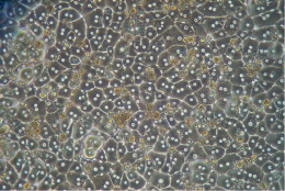
#1- Human Colonic Epithelial Primary Cells Total Kit (HCnEpC)

The organization of the epithelium of the small intestine and colon into crypts is generally comparable.
However, histologically there are two important differences between the two types of epithelia. The colonic epithelium has a flat epithelium surface and does not have villi. Regeneration occurs from units embedded within the crypts, each containing about 6–10 stem cells, while differentiated epithelial cells reside at the surface. Human colonic epithelium is linked to various human diseases and disorders such as Ulcerative Colitis and Colon Cancer, necessitating a better understanding of epithelial cell function and their role in disease-affected tissues.
Cell Application/tebu-bio’s colonic epithelial culture system provides a unique tool for the investigation of colon epithelial cell physiology and evaluation of therapeutic mediators.
-
Human Colonic Epithelial Cells Total Kit (Cat. nr 732CnK-10f or 732CnK-20f)
#2- Human Intestinal Epithelial Cells Total Kit (HInEpC)

The intestinal epithelium is a single layer of cells organized into crypts and villi, known as the most rapidly self-renewing tissue in adult mammals. The cells that line the intestinal lumen perform the primary functions of digestion, water and nutrient absorption, and forms a barrier against luminal pathogens.
Cell Applications / tebu-bio’ intestinal epithelial culture system provides outstanding resource for investigation of intestinal epithelial cell physiology related to GI infection, inflammatory bowel disease (IBD) like Crohn’s disease, ulcerative colitis, and intestinal cancer.
This epithelial cell culture system can be efficiently used as a test platform for the potential drug candidates and disease modulators. Other applications include functional analysis of intestinal epithelium, GI disease modeling, preclinical testing, drug compound screening and other validation assays.
-
Human Intestinal Epithelial Cells Total Kit (Cat. nr 732In-10f or 732In-20f)
#3- Human Gastric Epithelial Cells (HGaEpC)

The gastric mucosa is the mucous membrane layer of the stomach lumen which contains the glands and the gastric pits.
The human stomach lumen is lined with a monolayer of epithelial cells. The gastric pathogen Helicobacter pylori is one of the most common pathogens to colonize the stomach, and chronic infection can lead to gastric ulcers, gastritis, and gastric cancer.
Cell Applications/tebu-bio provide a gastric epithelial culture system that results in a gastric epithelial cell monolayer with tight junction that is stable for up to a week in culture enabling effective research of gastric diseases and disorders in vitro.
These cells provide researchers a valuable in vitro model for investigation of gastric epithelial cell physiology and their response to infection, tight junction function, as well as testing of therapeutic agents for gastrointestinal research.
-
Human Gastric Epithelial Cells Total Kit (Cat. nr 732GaK-10f or 732GaK-20f)
Looking for other Epithelial primary cell types to amplify your cell cultures ?
Cell Applications is going to release validated Epithelial cells from various tissular sections of the Human GI tract (eg. duodenum, jejunum, ileum). If you are looking for particular tissues, don’t hesitate to contact me via the form below.
Research laboratories and screening platforms sometimes require large numbers of mammalian cells. Therefore, cell culture capacity and timely supply of cells are challenges you might face regularly. In order to assist you optimally, tebu-bio’s laboratories have developed a series of convenient services which could be very useful to you:
- Primary cells & cell lines amplification – click here to know more about these services.
- 3D cell culture spheroid production lab services as described here.
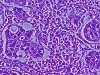
Recently, tebu-bio’s scientists used the HCT-116 cell line to produce a batch of colon carcinoma spheroids. Cells were given the conditions to form a cohesive and organized tissue. Spheroids capture epithelial tissue traits and respond to drugs and to 3D matrix environments in a close to in vivo manner. Colorectal carcinoma cell line HCT-116 spheroids were then treated with paclitaxel. In parallel, Liver carcinoma cell line HepG2 spheroids were treated with staurosporine. The dose-dependent effect of paclitaxel and staurosporine on spheroid cell viability was determined with the CCK-8 colorimetric assay. These results published for paclitaxel (IC50= 4,03 and 9,5nM – Sirenko et al., 2015), and for staurosporine (IC50 =8,9±0,4 – Sirenko et al., 2015) were within the range of previously published data.
You might like to download the results of these PoC tests here : HCT-116 Spheroids with paclitaxel & HepG2 spheroids with staurosporine.
Sources:
- Kang D.W. et al. “Reduced incidence of Prevotella and other fermenters in intestinal microflora of autistic children.” PLOS One 8, e68322 (2013)
- Scheperjans F. et al. “Gut microbiota are related to Parkinson’s disease and clinical phenotype”. Movement Disorders, 2015 Mar;30(3):350-8.
- Sirenko o. et al. “High-Content Assays for Characterizing the Viability and Morphology of 3D Cancer Spheroid Cultures” Assay Drug Development Technologies, 2015 Sep 1; 13(7): 402–414.
- Keshavarzian A. et al. “Colonic bacterial composition in Parkinson’s disease” Movement Disorders, 2015 Sep;30(10):1351-60.
- Unger M.M. et al. “Short chain fatty acids and gut microbiota differ between patients with Parkinson’s disease and age-matched controls”. Parkinsonism Related Disord. 2016 Nov;32:66-72.
- Kang D.W. et al. “Microbiota Transfer Therapy alters gut ecosystem and improves gastrointestinal and autism symptoms: an open-label study.” Microbiome. 2017 Jan 23;5(1):10
- Rumblings of Parkinson’s: Gut Microbiome Shifts in Early Stage of Disease – Alzforum (April 2017)
- Yuhai Z. et al. “Microbiome-Derived Lipopolysaccharide Enriched in the Perinuclear Region of Alzheimer’s Disease Brain” Frontiers in Immunology, 2017, 8:1064