In previous posts, I invited you to look at versatile methods to
- Visualize actin: Focus on Actin staining and visualization
- Measure actin binding: Focus on Actin – Detection of actin binding and actin binding proteins
- Measure actin polymerization: Focus on Actin – Measuring actin polymerization

Today, let’s focus on a method which enables researchers to measure the ratio between monomeric globular actin (G actin) and filamentous actin (F actin) in cells, thus giving a good insight in the status of actin dynamics.
The actin cytoskeleton is critical to many cellular functions, including the maintenance and regulation of cell shape, motility, intracellular communication, intracellular transport and the transduction of extracellular signals into the cell. Understanding the mechanisms that directly and indirectly regulate the actin cytoskeleton is an important area of research, and quantitation of the G-actin / F-actin ratio is a useful metric in helping define these mechanisms.
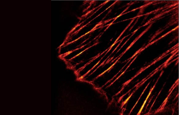
G actin readily polymerizes under physiological conditions to form F actin which is a double-helical filament (Fig. 1). Actin can polymerize from both ends in vitro. However, the rate of polymerization is not equal. This results in an intrinsic polarity in the actin filament. It has therefore become the convention to term the rapidly polymerizing end the plus-end or barbed-end (+) while the slow growing end is called the minus-end or pointed-end (-).
Measuring G:F actin ratio in cells

The most reproducible and accurate method of determining the amount of F actin content versus free G actin content in a cell population is to use Western blot quantitation of F actin and G actin cellular fractions. The general approach is to homogenize cells in F-actin stabilization buffer, followed by centrifugation to separate the F-actin from G-actin pool. The fractions are then separated by SDS-PAGE and actin is quantitated by Western blot. The final result gives the most accurate method of determining the ratio of F-actin incorporated into the cytoskeleton versus the G-actin found in the cytosol.
Cytoskeleton Inc. offers this method in an easy-to-handle and ready-to-use G-Actin : F-Actin Assay Kit. Typical results obtained with this kit are shown in Fig. 2. Changes in the amount of G-actin and F-actin were investigated in Swiss 3T3 cells treated with the actin polymerizing drug jasplakinolide, using the G-actin/F-actin in vivo assay kit. In untreated Swiss 3T3 cells, 80% of actin is soluble G-actin, and is found within the supernatant fraction, 20% of actin is filamentous F-actin and is found in the pellet fraction. In Swiss 3T3 cells treated with jasplakinolide, 80% of actin is reorganized into F-actin and is found in the pellet fractio.
The kit contains comes with all reagents needed to perform this assay and contains sufficient materials for 30-100 assays depending assay setup and includes reagents for positive and negative controls.
If you’d like an overview about the complete offer available in the actin field, take a minute to browse this Actin Product Guide.
Interested? Please contact me through the form below to get further information.