Research nowadays aims at working on models as similar as possible to the real physiological status. This includes the modification of cell culture conditions, For example, one should perform cell culture under “real” oxygen levels (e.g. hypoxia, normoxia, physioxia). For circulating cells, shear stress is a key factor, as cells behave in a different way depending on whether they are cultured under static or dynamic conditions.
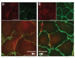
A publication by Melchior & Frangos (1) is a clear proof of this. They studied the differential phosphorylation of Akt-1 in endothelial cells depending on culture conditions. They found that the subcellular distribution of flow-induced Akt-1 phosphorylation in endothelial cells lining the mouse aorta varies depending on local hemodynamics. Activated Akt-1 accumulated in perinuclear areas of cells in regions predisposed to disturbed flow, but were localised at the cell-cell junction in regions of high unidirectional laminar shear stress.
This shows that Akt-1 signaling pathway is dynamically responsive to flow direction, thus offering a novel approach to regulating endothelial cell dysfunctions in regions subjected to flow reversal, as it happens in atherosclerosis.
Reference:
(1) Distinctive Subcellular Akt-1 Responses to Shear Stress in Endothelial Cells