Nowadays in Cancer research, there is a strong need for three-dimensional (3D) in vitro experimental approaches that mimick as much as possible the in vivo tumour ecosystem. In addition to solving issues related to animal models, these in vitro 3D models allow precise tuning of experimental design when investigating cancer cell biology but also, compound screening and disease modelling. In this post, we’ll take a look at the growth of the popular breast cancer cell line, MCF-7, using Alvetex®Scaffold technology (Reinnervate – a ReproCell company) to create a 3D structure that closely resembles the structure of tumour tissues grown in vivo.
Certain aspects of cancer research may rely on animal models, but associated with these are both feasibility and ethical concerns [1;2]. These issues may be overcome using in vitro cellular models. The vast majority of such in vitro models involve culturing cells on conventional two-dimensional (2D) plastic-ware, in which cells adapt to the flat polystyrene substrate, flatten, and grow as monolayers. Unfortunately, this approach is a poor surrogate and does not mimic the environment that cells experience in vivo. The morphology of cultured cells as monolayers isn’t realistic, cell-to-cell contact is limited and the microenvironment generated by the cells, for example via ExtraCellular Matrix (ECM) deposition, is reduced and altered [3].
Three-dimensional (3D) in vitro models are becoming a popular alternative to bridge and reduce the gap between 2D culture assays and animal models. Such 3D systems represent a more realistic approach enabling cells to retain their native 3D morphology, form interactions with adjacent cells and create more complex structures.
The Alvetex®Scaffold provides the ideal environment for cells to grow and function in 3D creating a more representative tissue model. The material consists of a highly porous and inert polystyrene scaffold with multiple voids and interconnects, creating a 3D environment which cells can freely grow in and occupy, creating 3D structures in vitro.
In this post, we will review the use of the Alvetex®Scaffold technology to create a 3D structure with the MCF-7 breast cancer cell line and compare its performance versus conventional 2D plastic-ware.
#1. Growth and Viability of MCF-7 cells on Alvetex®Scaffold
The analysis of the growth and expansion of MCF-7 cells cultured on Alvetex®Scaffold was assessed over 21 days.
MCF-7 cell viability was measured by the MTT assay and results showed good linear growth and expansion of the cell population during the first 2 weeks (Figure 1). Between 14 and 21 days, cell proliferation had plateaued and cells had reached confluence in the 3D construct.
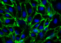
Histological data confirmed the increase of cell density over time (Figure 2). The penetration of the cells into the scaffold started from the top of the scaffold membrane where the cells were initially seeded. By day 7, the MCF-7 cells had reached the bottom of the membrane and continued to expand by forming aggregates of cells within the voids of the scaffold. Beyond 14-18 days, the cells had started to become overcrowded and confluent in 3D, and the quality of the culture was reduced resulting in areas of dead or dying cells.


Previous work has shown that MCF-7 cells transplanted into mice resulted in a xenograft tumour (Figure 3A). In the xenograft, MCF-7 cells grow as aggregates amongst a network of collagen fibres and diffuse connective tissues.
Similarly, MCF-7 cells cultured in Alvetex®Scaffold for 14 days formed aggregates as seen at higher magnification (Figure 3B). Furthermore, Masson’s Trichrome staining showed collagen deposits secreted by the cells indicating the production of ECM in the culture (Figure 3C).

Taken together, these data indicate that MCF-7 cells grown on Alvetex®Scaffold produce tissue-like morphologies resembling the structure of MCF-7 derived xenograft tissues.
# 2. Assessment of MCF-7 cytotoxicity to known compounds in 2D and 3D cultures
Cytotoxicity assays have been performed to compare the behaviour of MCF-7 cells grown either in 3D on Alvetex®Scaffold or 2D on conventional plastic-ware.
Three cytotoxic drugs were chosen based on their popularity in published toxicity studies. These drugs have been routinely used to study cancer cells in vitro and in animal models in vivo. These drugs are known to have human therapeutic applications.
- Tamoxifen is used in the treatment of estrogen receptor (ER)-positive breast cancer patients and as chemoprevention in high-risk women [4]. Tamoxifen is an antagonist of the estrogen receptor in breast tissue that decreases DNA synthesis.
- Paclitaxel is considered a highly active chemotherapeutic agent in various cancers including breast cancer [5]. Paclitaxel is a mitotic inhibitor that blocks the progression of mitosis.
- Doxorubicin is frequently used as a chemotherapeutic agent against metastatic breast cancers [6]. Doxorubicin is an anthracycline antibiotic, which works by intercalating DNA and thereby stopping the process of replication.
The assessment of drug cytotoxicity is normally performed on cells seeded on 2D plastic-ware that are usually exposed to the compound for 24h or 72h at a range of increasing concentrations in order to create a dose-response curve and to determine the IC50 value. The IC50 is the concentration of the compound required to kill 50% of the cells.
In an initial short term study, MCF-7 cells were grown for 3 days in 2D culture using conventional plastic-ware or in 3D culture using Alvetex®Scaffold. The cultures were subsequently treated with test compounds for either 24h or 72h. Figure 4 shows the growth curves obtained with Tamoxifen treatments. After both 24h and 72h treatments the viability of the cells was reduced to 100% mortality as the drug concentration increased in both 2D and 3D culture. However, the profiles of the responses were not the same for 2D and 3D cultures and the IC50 values were different. For 24h treatments the IC50 values were 12 µM and 32 µM for 2D culture and 3D culture, respectively. As would be anticipated, the effects of the drug were more marked after 72h incubation and IC50 values were 9 µM and 20 µM for 2D culture and 3D culture, respectively. For 2D cultures, these IC50 values are in agreement with those previously reported [7].

Histological analysis of MCF-7 cells treated for 24h with Tamoxifen confirmed the viability assay results. As the drug concentration increased, the cell morphology changed and the quantity of cells within the scaffold decreased. In control (0 µM Tamoxifen) cultures, cells possessed a large clear cytoplasm and close inter- cellular connections with adjacent cells. As the concentration of Tamoxifen increased, the cells became rounded, smaller and died, and some were lost floating into the medium.
These results indicate that MCF-7 cells cultured on Alvetex®Scaffold are more robust and that they can tolerate a higher concentration of Tamoxifen. In contrast, cells grown in 2D culture are placed under greater stress due to their abnormal structure and are therefore likely to be more sensitive when challenged by this compound.
Cytotoxicity assays have been performed to compare the behaviour of MCF-7 cells grown either in 3D on Alvetex®Scaffold or 2D on conventional plastic-ware.
Similar results were obtained with Paclitaxel treatment (Figure 5A). After 24h, 40% of the cells grown in 2D showed cytotoxicity toward this compound whereas only 20% of cells grown on Alvetex®Scaffold were no longer viable. A similar profile was observed after 72h treatment (data not shown). It is known that MCF-7 cells from ECACC are partially resistant to Paclitaxel [8] and such resistance appears to be greater in 3D culture compared to 2D culture.
Responses to Doxorubicin after 72h exposure were similar between cells cultured in 2D on conventional plastic-ware and in 3D on Alvetex®Scaffold (Figure 5B). The IC50 values were 1 µM and 0.7 µM for 2D culture and 3D culture, respectively. The 2D IC50 value is in agreement with previous work [7]. Interestingly, a small proportion of cells grown on Alvetex®Scaffold in 3D culture appear to be resistant at the higher drug concentrations. Whilst 100% cell mortality can be achieved with 4 µM Doxorubicin in 2D culture, between 5-10% of the cell population remains viable when grown on Alvetex®Scaffold. This example demonstrates how resistance to a compound drug could be overlooked if the cells were responding in an aberrant way due to growth in 2D culture.

To more closely mimic the treatment and response of tumour tissues to cytotoxic compounds, experiments were performed on more established 3D cultures of MCF-7 cells.

MCF-7 3D cultures were left to grow for 10 days prior to drug treatment. Under these conditions, the MCF-7 cells form much denser 3D tissue-like masses that resemble the structure of xenograft tumours (Figure 3). MCF-7 10 day 3D cultures were then exposed to Tamoxifen or Doxorubicin at concentrations based approximately on their IC50 values determined from 2D cultures after 72h treatment (10 µM and 1 µM, respectively). Figure 6 shows data relating to the cytotoxicity of these compounds on MCF-7 cells grown in 3D culture for 10 days followed by drug exposure for up to 18 days.
During the first five days of treatment with either Tamoxifen or Doxorubicin a significant delay in the response of MCF-7 cells grown in Alvetex®Scaffold was identified compared to conventional 2D cultures. Indeed for the cells grown in 2D plastic-ware the cytotoxicity of the compounds was effective from the first days of treatment. After 10 days of exposure, the proportion of cells responding to either compound was similar for cells grown in 2D or 3D culture. However, although no viable cells were detected in 2D cultures after 10-14 days, a small proportion of the 3D MCF-7 cultures remained viable for up to 18 days of treatment with either Tamoxifen or Doxorubicin. It is possible that a fraction of the cell population in 3D culture was resistant to the compounds under these test conditions but no such cells were found in 2D culture. Taken together, these results further indicate that MCF-7 cells cultured in 3D on Alvetex®Scaffold are more robust and that resistance to candidate molecules is observed which could be overlooked if the cells were grown in 2D culture.
#3 . Compatibility of Alvetex®Scaffold formats for cancer cell cytotoxicity assays
Alvetex®Scaffold is available in well insert and multi-well plate formats.
The well insert format is ideal for long-term cultures as cells receive nutrient support from above and below the 3D culture resulting in the formation of tissue-like structures. For example, in this study it has been proven that the formation of MCF-7- derived xenograft-like tumour masses in Alvetex®Scaffold presented in the 6-well insert format. Such growth is also possible in the smaller 12-well insert format. However, the use of well inserts and the need for long-term cultures may not be appropriate for higher throughput, short-term culture studies.
Accordingly, we have also demonstrated the application of MCF-7 3D culture and analysis of Tamoxifen cytotoxicity using 15 mm diameter Alvetex®Scaffold discs presented in the base of conventional 24-well plates. The appropriate cell seeding density and ratio of medium volumes have been calculated to enable comparison of cell growth and cytotoxic responses with the well insert format. Cells were grown for 3 days as before, prior to treatment with Tamoxifen (10-40 µM) for 24h or 72h. Experiments show that similar results can be obtained for short-term 3D culture of MCF-7 cells in either insert or plate formats of Alvetex®Scaffold. Consistent with earlier data performed using well inserts (Figure 4), treatment of MCF-7 cells in 3D culture in 24-well plates with Tamoxifen for either 24h or 72h reduced cell viability in a concentration dependent manner. The IC50 values generated from using either inserts or plates were highly comparable: 24h treatment – 32 µM and 30 µM; 72h treatment – 20 µM and 24 µM, respectively. These data indicate the compatibility of alternative presentations of Alvetex®Scaffold technology which is valuable for comparative studies in alternative formats and extrapolation into high throughput studies when using the 96-well plate presentation.
Benefits of Alvetex® Scaffold in 3D cell culture
Altogether, data shows that MCF-7 cells cultured on Alvetex® Scaffold possess a different sensitivity pattern to certain cytotoxic drugs than their 2D counterparts that is more likely to resemble in vivo responses.
This highlights the value of the Alvetex®Scaffold technology by providing a superior culture environment for cancer cell research.
This study demonstrates that human MCF-7 breast cancer cells grown in Alvetex®Scaffold provide a realistic model that is a step closer to replicating the in vivo-like conditions of tumour growth. MCF-7 cells show a linear growth profile on Alvetex®Scaffold for 2 weeks before they reach confluency and plateau. The 3D morphology of the cells, the opportunity for close interactions between adjacent 3D cells, and the production of extracellular matrix within Alvetex®Scaffold contribute toward creating tissue-like structures that closely resemble the morphology of xenograft tumours. The cytotoxicity assays performed on these 3D cultures generate valuable data concerning the sensitivity and toxicity of cells to specific compounds.
By avoiding the limitations of conventional 2D culture, Alvetex®Scaffold technology provides new opportunities to study the effectiveness and mode of action of test compounds on cancer cells. Creation and evaluation of xenograft-like structures in vitro is likely to generate more appropriate data concerning the predictive accuracy of a compound test assay compared to cells compromised by their growth conditions in 2D culture. Furthermore, such 3D models may partially substitute the need for animal experimentation hence reducing animal usage and associated costs.
Overall, Alvetex®Scaffold provides a simple and robust approach for the routine generation of 3D culture models to more accurately study the action of test compounds in cancer research.
Sources
- Knight, A., Systematic reviews of animal experiments demonstrate poor human clinical and toxicological utility. Altern Lab Anim, 2007. 35(6): p. 641-59.
- Wells, D.J., Animal welfare and the 3Rs in European biomedical research. Ann N Y Acad Sci, 2011. 1245: p. 14-6.
- Amada, K.M. et al., Modeling tissue morphogenesis and cancer in 3D. Cell, 2007. 130(4): p. 601-10.
- Fisher, B. al., Tamoxifen for the prevention of breast cancer: current status of the National Surgical Adjuvant Breast and Bowel Project P-1 study. J Natl Cancer Inst, 2005. 97(22): p. 1652-62.
- Saloustros, E., et al., Paclitaxel and docetaxel in the treatment of breast cancer. Expert Opin Pharmacother, 2008. 9(15): p. 2603-16.
- Moreno-Aspitia, A. et al., Treatment options for breast cancer resistant to anthracycline and taxane. Mayo Clin Proc, 2009. 84(6): p. 533-45.
- Panasci, L. al., Sensitization to doxorubicin resistance in breast cancer cell lines by tamoxifen and megestrol acetate. Biochem Pharmacol, 1996. 52(7): p. 1097-102.
- Ajabnoor, G.M. et al., Paclitaxel resistance is associated with switch from apoptotic to autophagic cell death in MCF-7 breast cancer cells. Cell Death Dis, 2012. 3: p. e260.