Antibody-based techniques are widely used in Life Science laboratories. Antibody purification is often required to raise purity yield of antibody production batches or to reach publication-grade data. In this post, take advantage of some technical tips and troubleshooting points to improve your antibody purification.
 Though most of the antibodies used in research are available in rather pure format, antibody purification is still regularly performed in research labs. This is normally achieved by using the well-known proteins A and G .
Proteins A and G are bacterial immunoglobin (IgG) binding proteins originating from Staphylococcus or Streptococcus respectively (protein A/G is a recombinant version coupling Protein A and G). Proteins A, G, and A/G are available in unconjugated and conjugated forms and optimized to run the most common immuno-assays (WB, ELISAs or IHC…).
Protein A, Protein G, A/G – which one should I choose?
In general, protein G is preferred, as it has “relatively” higher binding affinities. Nevertheless, the type of sample to purify and the type of applications are 2 important elements to keep in mind when designing your antibody purification. This is especially true if you are working with low affinity antibodies.
If you are looking for specific species, feel free to leave a message at the end of this post.
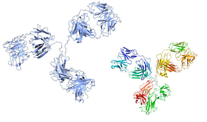
 Protein A

Protein A contains five homologous Ig-binding domains able to bind the Fc fragment of a wide range of mammalian immunoglobulins.
Protein A can be coupled to solid matrixes (ex. Sepharose™) for purification of Igs from ascites, serum or hybridoma culture media. When conjugated to a reporter tracer (ex. fluorescent dye, enzyme, biotin…, protein A is used to detect an Ig of interest (Dot blot, WB…). Protein A binding capacities might differ from sample species as described in the list you can download at the link above.
In this case, the precise selection of the Protein together with the flow used in your purification column are critic. You might interrupt of flow of liquid in your purification column so that the interaction between the antibody of interest and the protein conjugated to the resin is maximized.
 Popular Protein A related products and respective applications:
- Protein A Peroxidase Conjugate for ELISA and WB
- Protein A Biotin Conjugate for ELISA, WB and IHC
- Protein A Fluorescein Conjugate for FLISA and IF
Protein G
Protein G is an Ig-binding protein (see here for Protein G binding capacities). The current recombinant versions of Protein G have been optimized so that it does not bind anymore to albumin. Protein G is used for immunoglobin purification and detection and is also often immobilized on solid surface (ex. Sepharose™, latex or magnetic particles) for IgG purification from ascites, serum or hybridoma culture media. Protein G is also available in formats conjugated with fluorescent or enzymatic reporter molecules.
Popular Protein G related products and respective applications:
- Protein G for ELISA, WB and Ig purification
- Protein G Alkakine Phosphatase Conjugate for ELISA, WB and IHC
- Protein G Peroxidase Conjugate for ELISA and WB
Protein A and Protein G Sepharose™
Protein A, Protein G and Protein A/G Sepharose™ are available for high purity separation of monoclonal and polyclonal antibodies compatible with R&D and industrial scales. Rockland’s high quality Protein A, Protein G and Protein A/G Sepharose™ are fast flow, low leakage affinity resins. The coupling chemistry used minimizes leakage of Protein A or Protein G ligands and pampers IgG-binding activity with a long term stability from pH ranging to 3 to 9. For each lot, IgG-binding capacity is validated by human IgG binding assays to assure high quality of product.
Other elements to consider for optimal use of Protein A and Protein G
Optimized and fresh buffers, medium and resin are also a imortant for a successful purification.
- Avoid magnetic stirrers
- Use the buffers specific for each affinity medium / resin
- Make sure to control elution
- Adapt the pH. Low pH is needed to elute proteins from protein G. If this low pH (sometimes as low as pH 2.7) affects your antibody, use Protein A instead.
Sometimes, single-step purifications based on Fc region specificity will co-purify host IgG and trace amounts of serum proteins may be present in the final elution. In order to improve the purity levels, a multi-step purification is then recommended.
But what if I’m running WB? Any tips there?
Yes, there are some useful tips here too… WB is one of the most widely used techniques in laboratories worldwide. Protocols written by researchers are easily available and some really well describe the step-by-step WB procedure.
Nevertheless, experimental optimization is sometimes required to obtain publication-grade results (see tebu-bio’s tips for phosphorylated protein WB here).
Any questions? Don’t hesitate to leave your comments below.