Ubiquitin (Ub) is an 8 kDa highly conserved polypeptide, commonly expressed in eukaryotic cells. Ub is added to lysine residues of the target proteins. This post-translational modification (known as ubiquitination) is made through the sequential action of 3 enzymes (E1 Ub activating enzyme, E2 conjugating enzyme and E3 ligase).
In parallel, the 7 lysines of the Ub itself (K6, K11, K27, K29, K33, K48, and K63) are also Ub acceptors. This enables the formation of polyubiquitin chains among which, K48 and K63 linkages are the most studied. In addition to these 7 lysines, polyubiquitin linkages can be formed through a methionine residue present in the Ub polypeptide by the Linear Ubiquitin Chain Assembly Complex (LUBAC).
Ubiquination, a post-translational modification regulating various cell functions
Like phosphorylation or sumoylation, ubiquitination controls the activity and fate of the ubiquinated proteins and thus cellular functions. It is well accepted that:
- K48 Ub-protein linkages lead to proteasomal degradation.
- K63-Ub-protein linkages drive cell signaling, receptor endocytosis, cell trafficking and immune responses, the role of the linear and K6, K11, K27, K29, K33 Ub-protein not being completely understood.
- The linear polyubiquitin (M1-linked polyubiquitin) plays important roles in inflammatory and immune responses, by interacting with the NFKB signaling pathway.
3 monoclonals to discriminate Ub and polyubiquitin chains
Being able to distinguish the various forms of Ub present in a sample is very significant. It allows researchers to improve their knowledge of protein and cellular functions involved in the model analyzed. For such comparative studies, antibodies that recognize and distinguish linear polyubiquitin from polyubiquitylated proteins and free ubiquitin are necessary. Here, you will find my selection of highly qualified monoclonal antibodies to enable experimental approaches that will allow you to differentiate and localise the various UB forms in your samples;
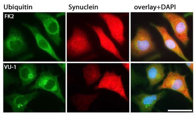
#1 – VU-1 monoclonal anti-Ubiquitin
VU-1 is a monoclonal antibody that recognizes K48-, K63-, K11- chains and linear free ubiquitin.
VU-1 is ideal for Western blotting (recommended dilution 1:1,000 – 1:10,000), immunostaining (recommended dilution 1:200 – 1:500) and ELISA.
#2 – FK2 monoclonal anti-Ubiquitin
The FK2 monoclonal ubiquitin antibody (AB120) detects both mono- and K11-, K29-, K48- and K63-linked polyubiquitin but not free ubiquitin! FK2 is suited for Western blotting (recommended dilution 1:1,000), immuno-precipitation and immunostaining applications.
#3 – LUB9 monoclonal anti-Ubiquitin
The Linear Polyubiquitin specific monoclonal LUB9 antibody (AB130) displays high specificity for linear polyubiquitin chains.
LUB9 is suitable for Western blotting of linear ubiquitin chains from tissue or cell lysates (recommended dilution 1:1,000 – 1:2,500). LUB9 specifically detects linear over K11-, K48- and K63-linked polyubiquitin.

Small molecules can be very useful to study this pathway. I have made a selection of small molecule active on the ubiquitin pathway for research and screening studies.
Feel free to suggest those you think should be added!
Want to discriminate linear- from poly- and free ubiquitin?
Think VU-1, FK2 and LUB9 monoclonal antibodies!
The experimental data described below speaks for itself…